- Product Listings
- Compare Products
- Calculators
- Product Tours
- Expert Reviews
- Our Experts
- Company Directory
- About Beye
- Contact Us
Ⓒ 2026 Beye.com. All rights reserved.
This content is intended for health care professionals and providers only. The information contained on Beye.com, including text, graphics, images, and interactive activities, is for informational purposes only, and is not intended to be a substitute for professional medical advice. Beye LLC, via its Editors and Publisher, accepts no responsibility for any injury or damage to persons or property occasioned through the implementation of any ideas or use of any product described herein. Although great care is taken to ensure that all information is accurate, it is recommended that readers seek independent verification of advice on drugs and other product usage, surgical techniques and clinical processes prior to their use. References made in article may indicate usage of medical equipment or drugs at dosages, for periods of time, and in combination not included in the current prescribing information. Inclusion of advertising materials on the website thereof, does not constitute and representation or guarantee by Beye LLC of the quality of such products, or of the claims made.
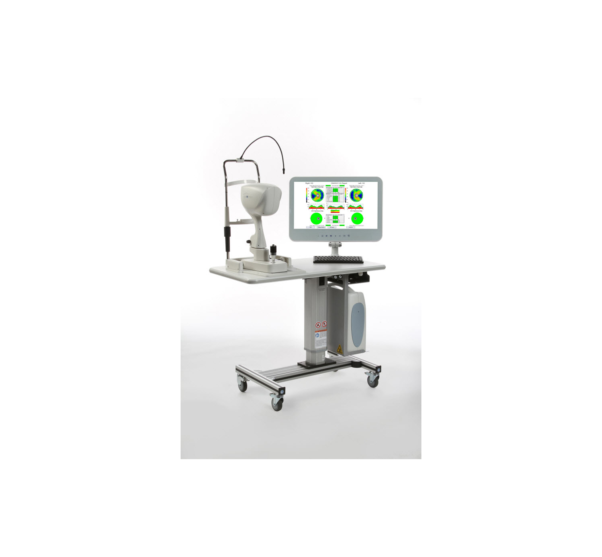
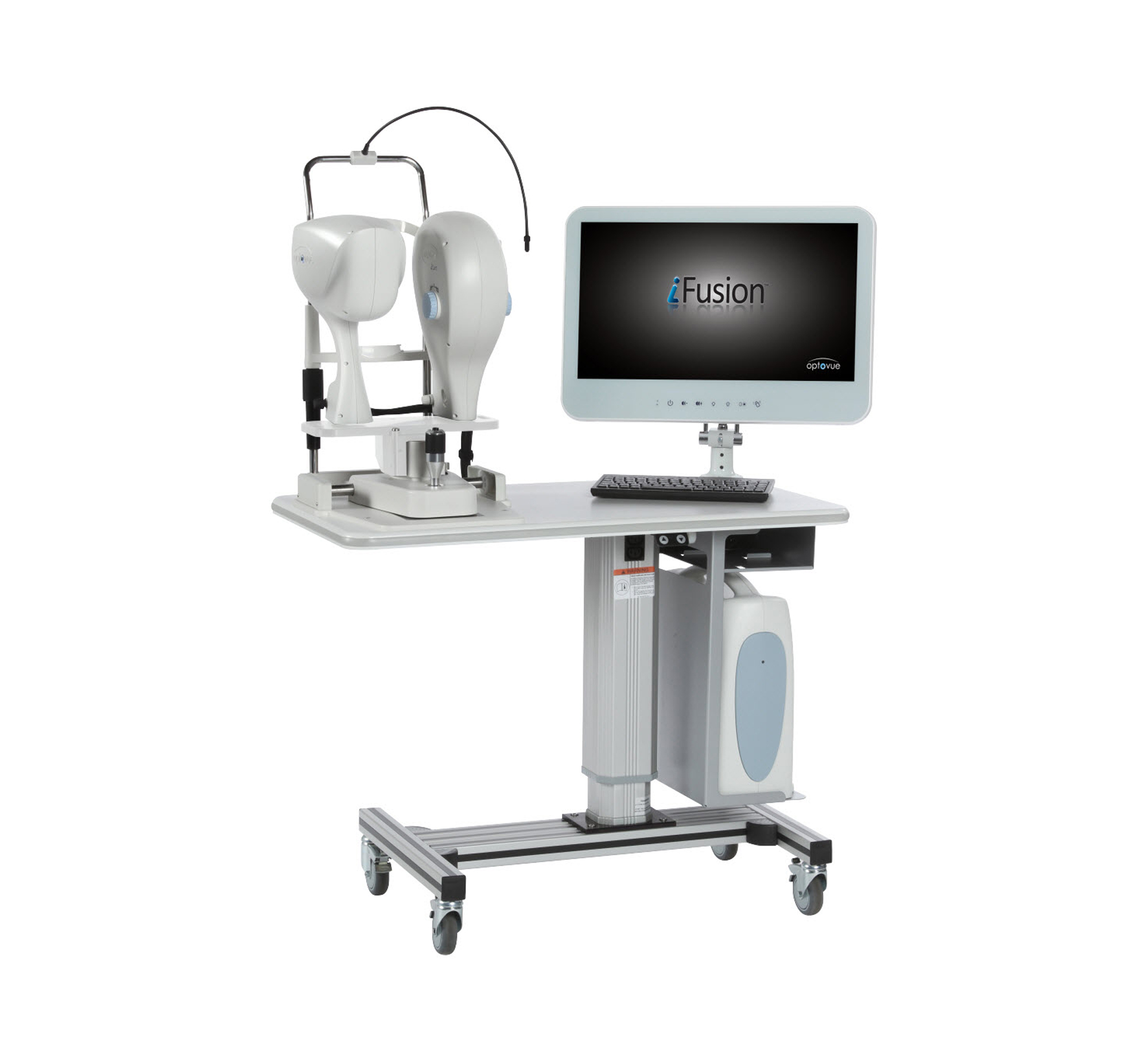
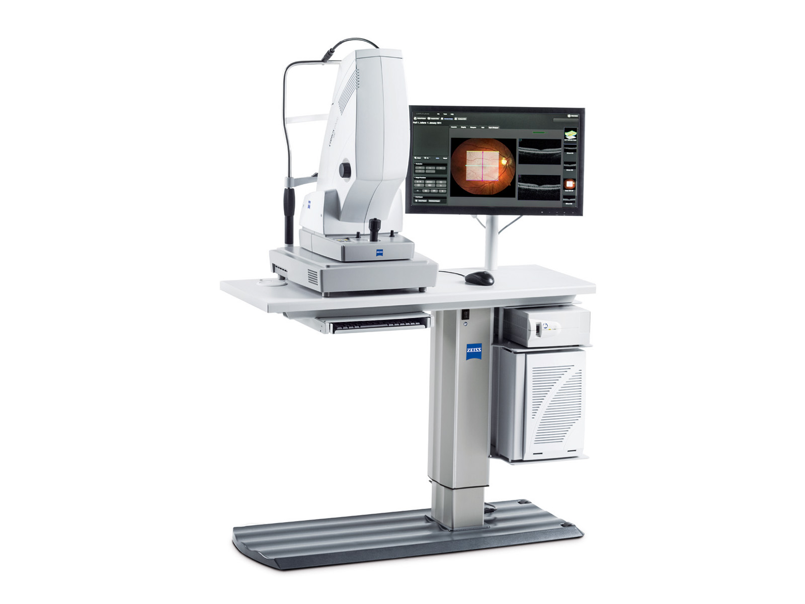
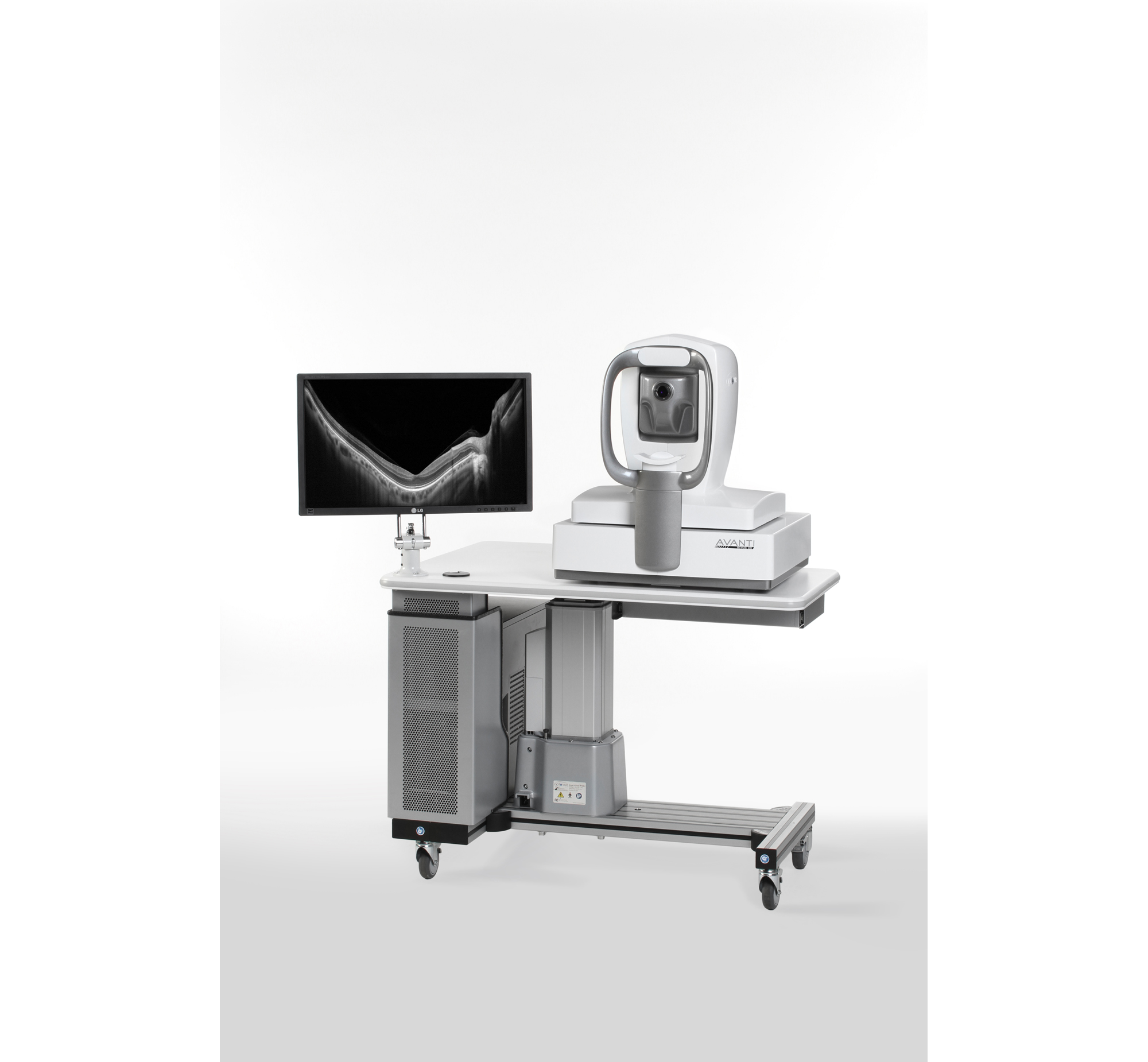
iVue
Optovue, Inc.iVue is the compact version of the RTVue OCT, offering the same scanning speed and resolution as the larger system, that includes scanning and reports for retina, retina nerve fiber and cornea assessment by the clinician. Epi-mapping is now available to provide corneal epithelia and stromal measurements that aid in the diagnosis, documentation, and management of ocular health.
At-a-Glance
- Virtual dissection of the retina and optic disc
- 512 x 128 dense cube with 67 million data points
- High density 3D volume for visualization and analysis of patient condition
Related Products

Details
FDA
Yes
CE Mark
Not specified
Capabilities
Not specified
Measurements
Normative Data Comparison, Optic Disc, RNFL Analysis, Ganglion Cell Complex (Optional)
Modes
Not specified
Processing
Optional 3D Reconstruction
User Ratings + Reviews
This is the newest piece of equipment we've added to our practice, and it has been very worthwhile. Excellent diagnostic tool, and right pricepoint for what we were looking for. It is advanced enough to get the numbers we need with good quality imaging. I still think CIRRUS is slightly better because it has a wider range, but in terms of what our practice needed, Optovue came in as something mid-range that we could do. The CIRRUS was more expensive.
Very easy to use, and great results. Nice to have different tests, testing anterior and posterior. I also like how the screen swivels so patients can easily see the results at testing, or when sent to exam rooms for viewing.
We obtained 3 of these for our practice in 2015. We also purchased the available Ganglion Cell Complex upgrade and 3D / en face analysis upgrade. We were allowed a generous trial period in the office and customer support and hands-on instruction are excellent. This includes anterior segment imaging for detailed looks at the chamber angle with excellent detail, as well corneal pachymetry mapping showing corneal thicknesses for at least 32 points on the cornea. The retinal imaging is exquisite, and retinal images can be viewed in an almost unlimited number of ways. For NFL and glaucoma evaluation, the images are excellent and much more reliable and accurate than our older non-spectral domain OCT. There is almost no signal drop-out even with very significant cataracts, and virtually no pupil dilation is necessary. It takes images very rapidly and takes up a very small footprint in the office. The techs picked up all aspects of using this machine very rapidly, and they love using it. It is fully networkable and compatible with EMR.
Show More
Company Information
Contact the company for additional information, availability, or pricing:
Optovue, Inc.
www.optovue.com2800 Bayview Drive
Fremont, CA 94538

Trending in Diagnostic Instruments
Powered by:
Tracking the Alignment of Toric IOLs
MillennialEye, May/June '18
Pseudophakic Bullous Keratopathy
William B. Trattler, MD
CRSToday, April 2018
Spotting AMD in the OD’s Office
CollaborativeEye, Mar/Apr '18
Spotting AMD in the OD’s Office
Nicole Stout, OD, FAAO, and Nate R. Lighthizer, OD, FAAO
CollaborativeEye, Mar/Apr '18
Talking Torics
CRSToday, March 2018
Cataract Surgery After LASIK Corneal Flap Removal
Karl G. Stonecipher, MD
CRSToday, March 2018
IOP After Short-Term Scleral Lens Wear in Healthy Adults
CollaborativeEye, Debut Issue
Ten Do’s and Don’ts for Cataract Comanagement
CollaborativeEye, Debut Issue
Persistent LASIK Flap Interface Fluid After DSAEK Procedure
CRSToday, February 2018
Persistent LASIK Flap Interface Fluid After DSAEK Procedure
Mark S. Gorovoy, MD
CRSToday, February 2018
Getting Another Shot
William B. Trattler, MD; Alan Berg, MD; and Naja Chisty, DO
CRSToday, January 2018
Dual Benefits of CXL
CRSTodayEurope, Mar 2013
Dual Benefits of CXL
CRSTodayEurope, Mar 2013
My Algorithm for DED
CRSToday, Jan 2016
Show More
