- Product Listings
- Compare Products
- Calculators
- Product Tours
- Expert Reviews
- Our Experts
- Company Directory
- About Beye
- Contact Us
Ⓒ 2026 Beye.com. All rights reserved.
This content is intended for health care professionals and providers only. The information contained on Beye.com, including text, graphics, images, and interactive activities, is for informational purposes only, and is not intended to be a substitute for professional medical advice. Beye LLC, via its Editors and Publisher, accepts no responsibility for any injury or damage to persons or property occasioned through the implementation of any ideas or use of any product described herein. Although great care is taken to ensure that all information is accurate, it is recommended that readers seek independent verification of advice on drugs and other product usage, surgical techniques and clinical processes prior to their use. References made in article may indicate usage of medical equipment or drugs at dosages, for periods of time, and in combination not included in the current prescribing information. Inclusion of advertising materials on the website thereof, does not constitute and representation or guarantee by Beye LLC of the quality of such products, or of the claims made.
Compare Spectral & Time-Domain OCTs
18 Products
reset all
At-a-Glance
Description
FDA
CE Mark
Capabilities
Image Capture
Measurements
Modes
Processing
Source
System
Type
Video Capture
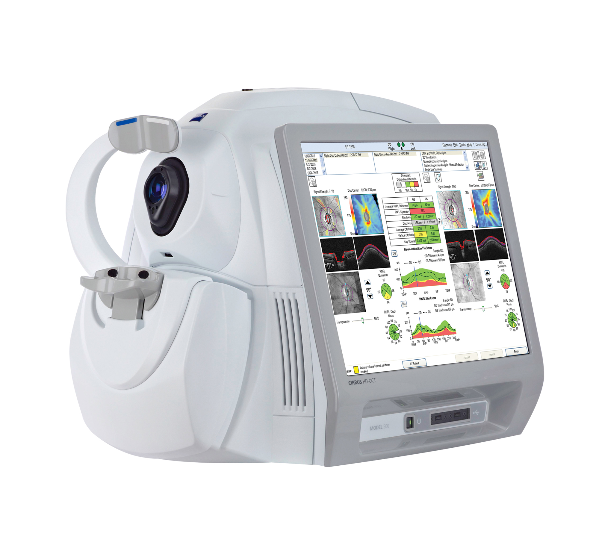
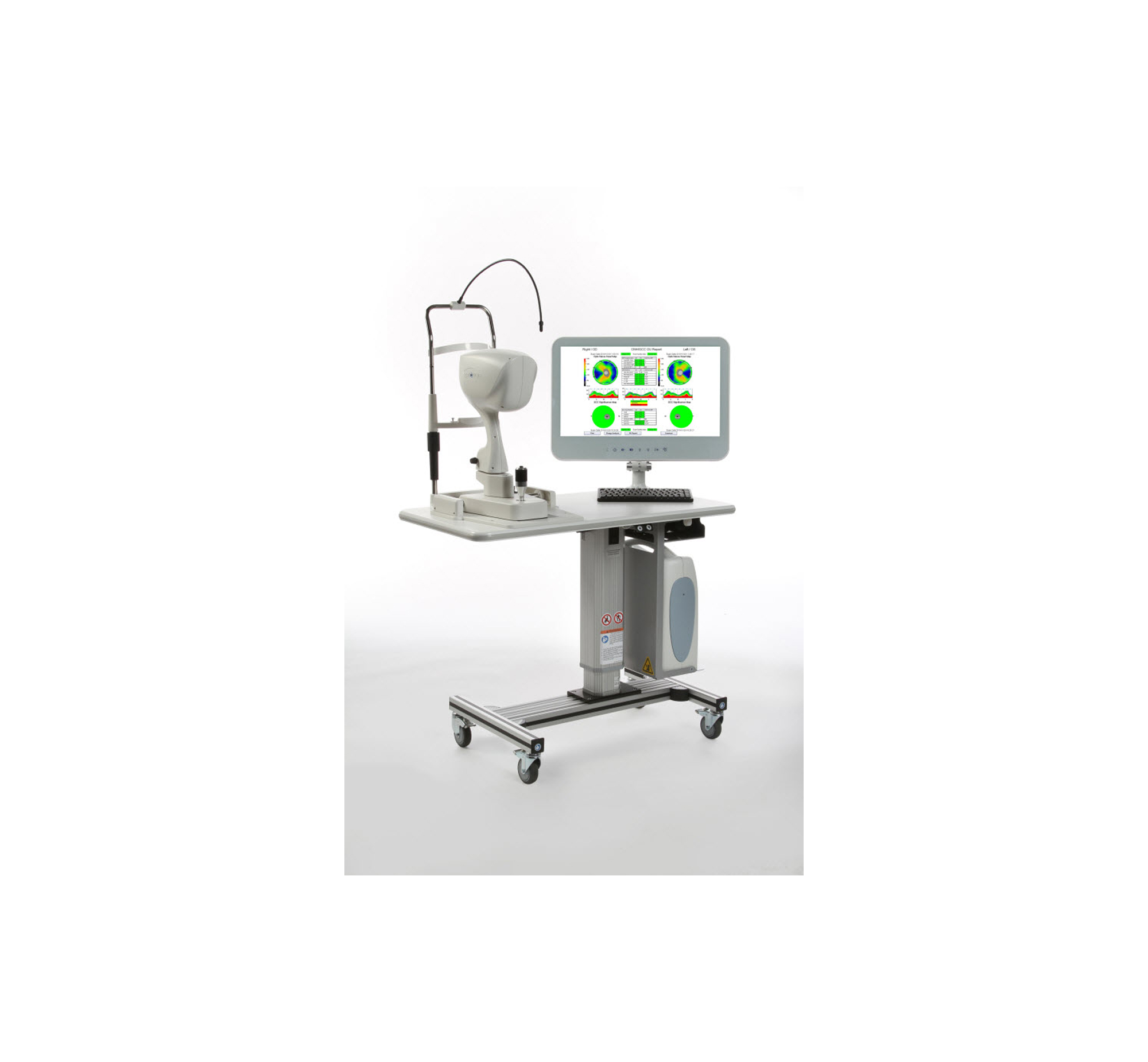
- Ganglion Cell Analysis
- Guided Progression Analysis (GPA)
- Macular Thickness and Change Analysis
- Macular Thickness Normative Data
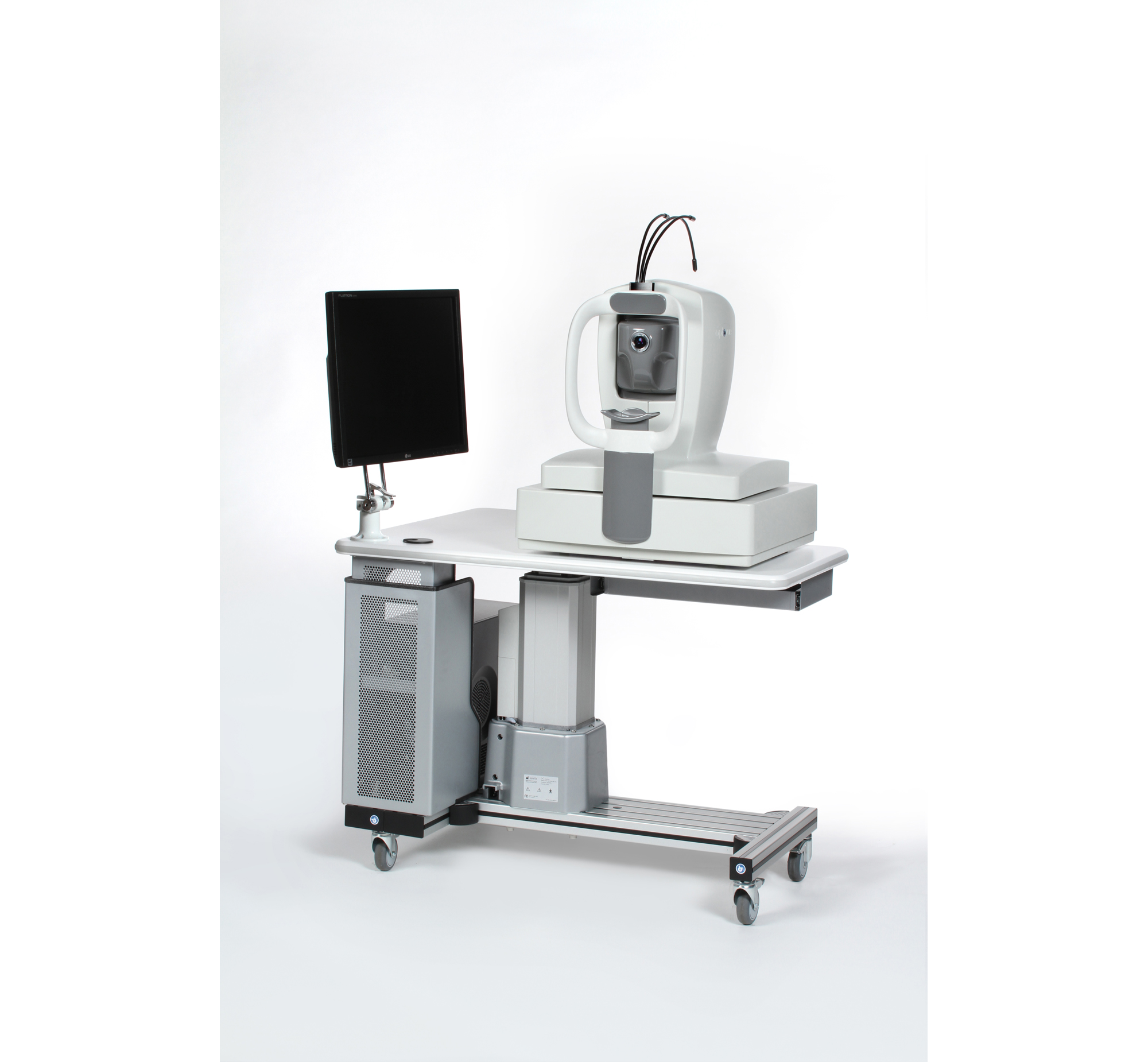
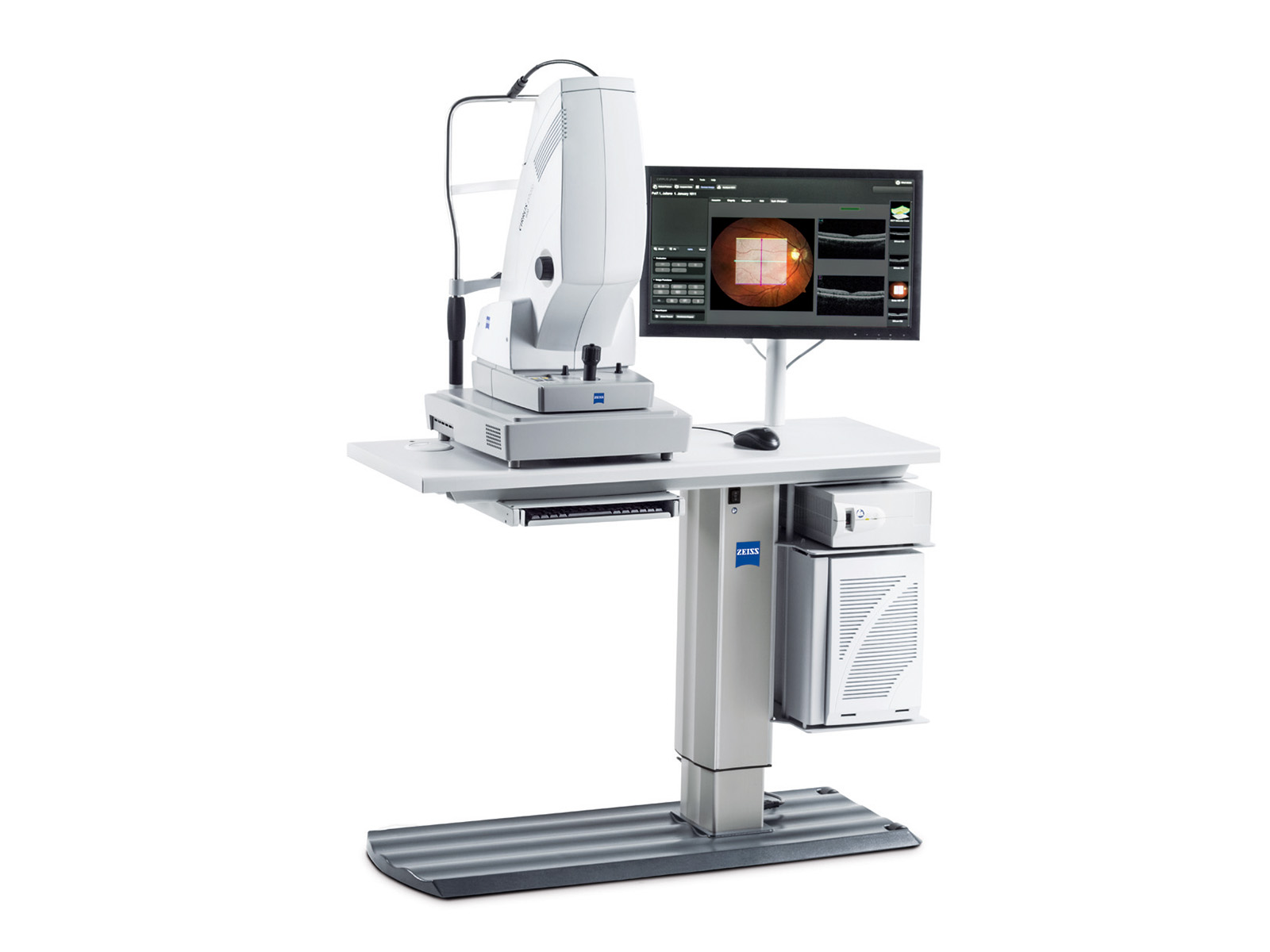
CIRRUS HD-OCT 500 provides a solution for comprehensive ophthalmic practices and offers essential OCT capabilities with a broad range of clinical applications in an easy-to-learn, easy-to-use instrument for the management of glaucoma and retinal disease, retina assessment for cataract surgery, and anterior segment imaging for corneal disease.
Yes
Yes
Retina Thickness, RPE Deformation, Corneal Thickness Mapping
—
Normative Data Comparison, RNFL Analysis, Ganglion Cell Complex
—
Not specified
—
Not specified
—
—
—
—
—
- Offers 1.6 μm Axial digital resolution in combination with the ability to average multiple scans
- The integrated Scanning Laser Ophthalmoscope (SLO) assists with follow-up examinations by automatically adjusting to the same scan position as used in the previous exam.
- To ease comparison, the software automatically selects identical scan parameters.
- Can automatically detect and distinguish 10 layers of the retina—including Bruch’s membrane (BM)
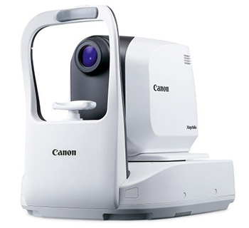
Xephilio OCT-A1 Optical Coherence Tomography device, together with the required RX Capture software, computer, and LCD monitor (collectively, the “Xephilio OCT-A1 System”), offers superb image quality. With an Axial digital resolution of 1.6 μm and a high scanning speed of 70,000 A-scans per second, the System enables excellent differentiation of structures and individual layers of the retina and can help with patient comfort.
Yes
Not specified
—
—
—
—
—
—
—
—
—

- Providing a comprehensive solution for retina and glaucoma analysis
- Accurate image capture with a SLO-based eye tracer
- Selectable OCT sensitivity that allows acquisition of B-scan images through media opacities
- Tracing HD for accurate averaging of up to 120 image
- Glaucoma analysis with wide-area normative database 9 x 9 mm
- High resolution AngioScan OCT-Angiography image
The RS-3000 Advance 2 incorporates a scanning laser ophthalmoscope and is designed for comprehensive imaging and analysis of the retina and glaucoma. Enhancements from the previous model include 85,000 A-scans/s, 12 x 12 mm auto panorama imaging of OCT angiography, and increased image quality.
No
Yes
Fundus Camera, Scanning Laser Imaging
Yes
OCT Angiography+Microperimetry, Normative DB+Microperimetry, Thickness Map DB+Microperimetry, Retinal mode, Choroidal mode
—
Not specified
—
Powercord
—
Not specified
—
—
—
- Fully automated operation with a simple finger touch
- Rich analysis and report functions
- Modular system with expandable features
- True color fundus photography
- Compact and space saving design
With a touch of the operation screen, the Maestro automatically scans both eyes and produces simultaneously an OCT scan & a true colour fundus image.
Yes
Yes
Fundus Camera, Wide-Field Imaging, Retina Thickness, Retinal Mapping, Corneal Thickness Mapping, Corneal Curvature Mapping
—
Scan Speed (Hz), A-Scan/Second, Scan Depth, Field Angle, Pupil Diameter, Normative Data Comparison, Optic Disc, RNFL Mapping, RNFL Analysis
—
Blue Reflectance (Red-Free), Scanning Laser Imaging
—
3D Reconstruction, Additional Image Processing Features
—
—
—
—
—
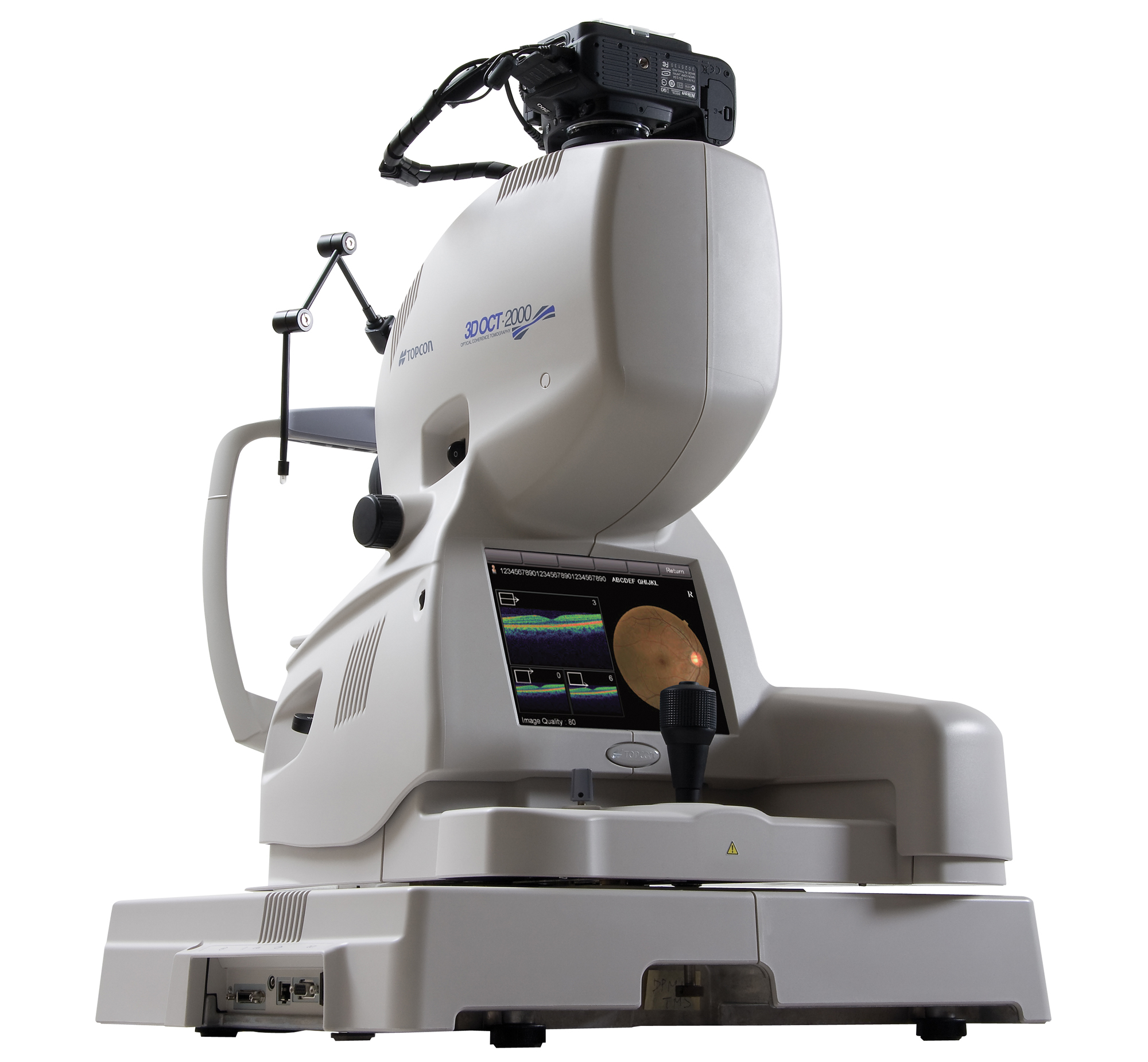
- Integrated, high resolution (12.3MP) fundus camera
- FastMap software enables dynamic viewing of 2D, 3D and fundus images simultaneously
- Embedded touch-screen for quick and easy navigation
- Historic patient data from Stratus OCT can be easily imported, analyzed and viewed
- Seamless integration with EyeRoute Image Management System
The 3D OCT 2000 series of spectral domain OCTs was designed to meet the needs of all eye care professionals from a single doctor practice to a large university hospital.
Yes
Not specified
Fundus Camera, Wide-Field Imaging, Retina Thickness, Retinal Mapping
—
Optic Disc, RNFL Analysis
—
ICG Angiography, Scanning Laser Imaging
—
3D Reconstruction
—
—
—
—
—
- Real-time active eye-tracking OCT
The RTVue Premier was the first FDA-cleared Spectral-Domain OCT launched in the United States, and also the first OCT cleared by the FDA for both corneal and retinal imaging.
Yes
Not specified
Not specified
—
Normative Data Comparison, Optic Disc, RNFL Analysis, Ganglion Cell Complex
—
Not specified
—
Not specified
—
—
—
—
—
- Virtual dissection of the retina and optic disc
- 512 x 128 dense cube with 67 million data points
- High density 3D volume for visualization and analysis of patient condition
iVue is the compact version of the RTVue OCT, offering the same scanning speed and resolution as the larger system, that includes scanning and reports for retina, retina nerve fiber and cornea assessment by the clinician. Epi-mapping is now available to provide corneal epithelia and stromal measurements that aid in the diagnosis, documentation, and management of ocular health.
Yes
Not specified
Not specified
—
Normative Data Comparison, Optic Disc, RNFL Analysis, Ganglion Cell Complex (Optional)
—
Not specified
—
Optional 3D Reconstruction
—
—
—
—
—
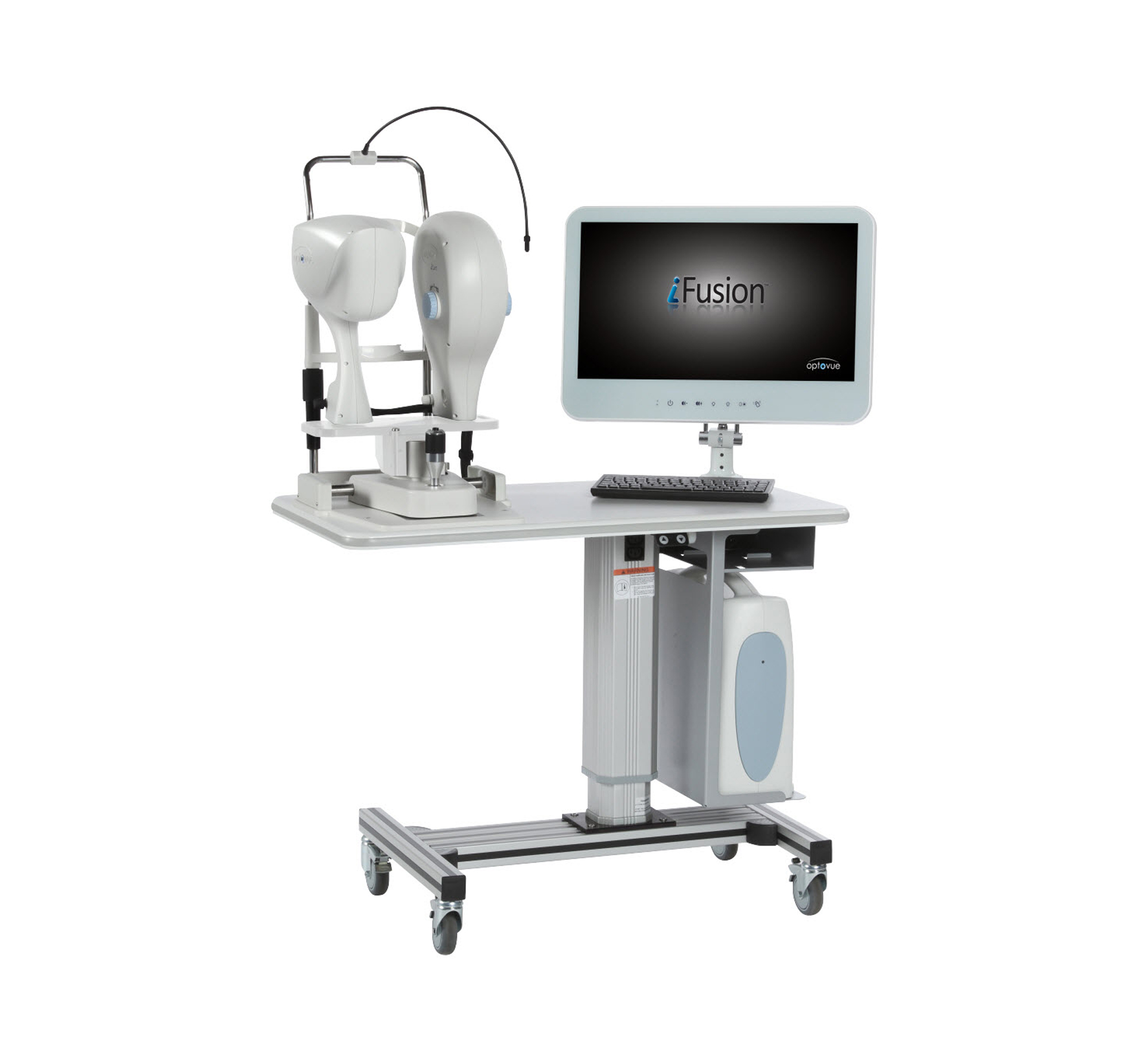
- Saves time & space for practice efficiency
- Upgrade options for more flexibility
iFusion combines Spectral-Domain OCT and Fundus imaging by adding the OCT capabilities of iVue and high quality imaging from iCam on a single, integrated, versatile platform for your practice. Epi-mapping is now available to provide corneal epithelia and stromal measurements that aid in the diagnosis, documentation, and management of ocular health.
Yes
Not specified
Not specified
—
Not specified
—
Not specified
—
Not specified
—
—
—
—
—
- Advanced RPE analysis
- Ganglion cell analysis
- Precision FoveaFinder
- Macular thickness and change analysis
- Macular thickness normative data
- Integrated 19 inch color flat panel display
CIRRUS HD-OCT 5000 is configured with high resolution visualization capabilities and sophisticated clinical applications such as Advanced RPE analysis to track retinal pigment epithelial integrity and Ganglion Cell Analysis. This helps to assess glaucomatous loss in the macula that may not be evident in the peripapillary region.
Yes
Yes
Retina Thickness, Corneal Thickness Mapping
—
Normative Data Comparison, RNFL Analysis
—
Not specified
—
Not specified
—
—
—
—
—
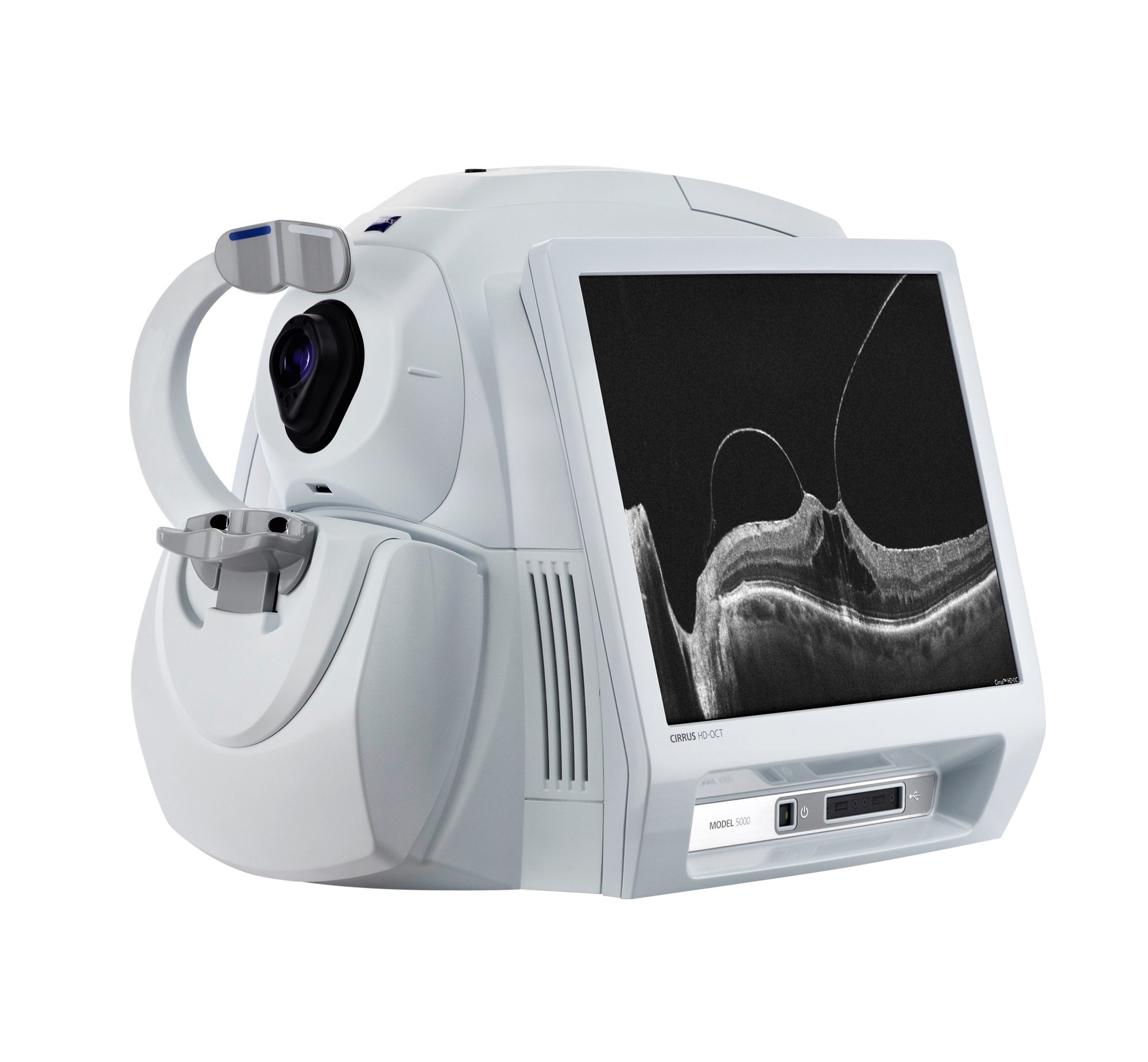
- Single-shot fundus autofluorescence
- Great detail density
- Interactive review
- Multimodal assessments
- Camera Modes: Autofluorescence, Infrared Reflectance, Blue Reflectance (red-free)
- Imaging Capabilities: Fundus camera, Retina thickness, Corneal thickness mapping
- Glauacoma Measurements: Normative Data Comparison, RNFL Analysis
- Size is 16.1 (W) x 18.9 (D) x 26.8 (H) in. and weight is 32.9 kg (72.7 lbs.)
CIRRUS photo combines a full mydriatic/non-mydriatic fundus camera with CIRRUS HD-OCT technology in one compact and versatile system. Available in two models, CIRRUS photo 600 and CIRRUS photo 800, it provides multiple insights for comprehensive retina and posterior segment care.
Yes
Yes
Fundus Camera, Retina Thickness, Corneal Thickness Mapping
—
Normative Data Comparison, RNFL Analysis
—
Autofluorescence, Infrared Reflectance, Blue Reflectance (Red-Free)
—
Not specified
—
Standalone
—
Mydriatic
—
—
—
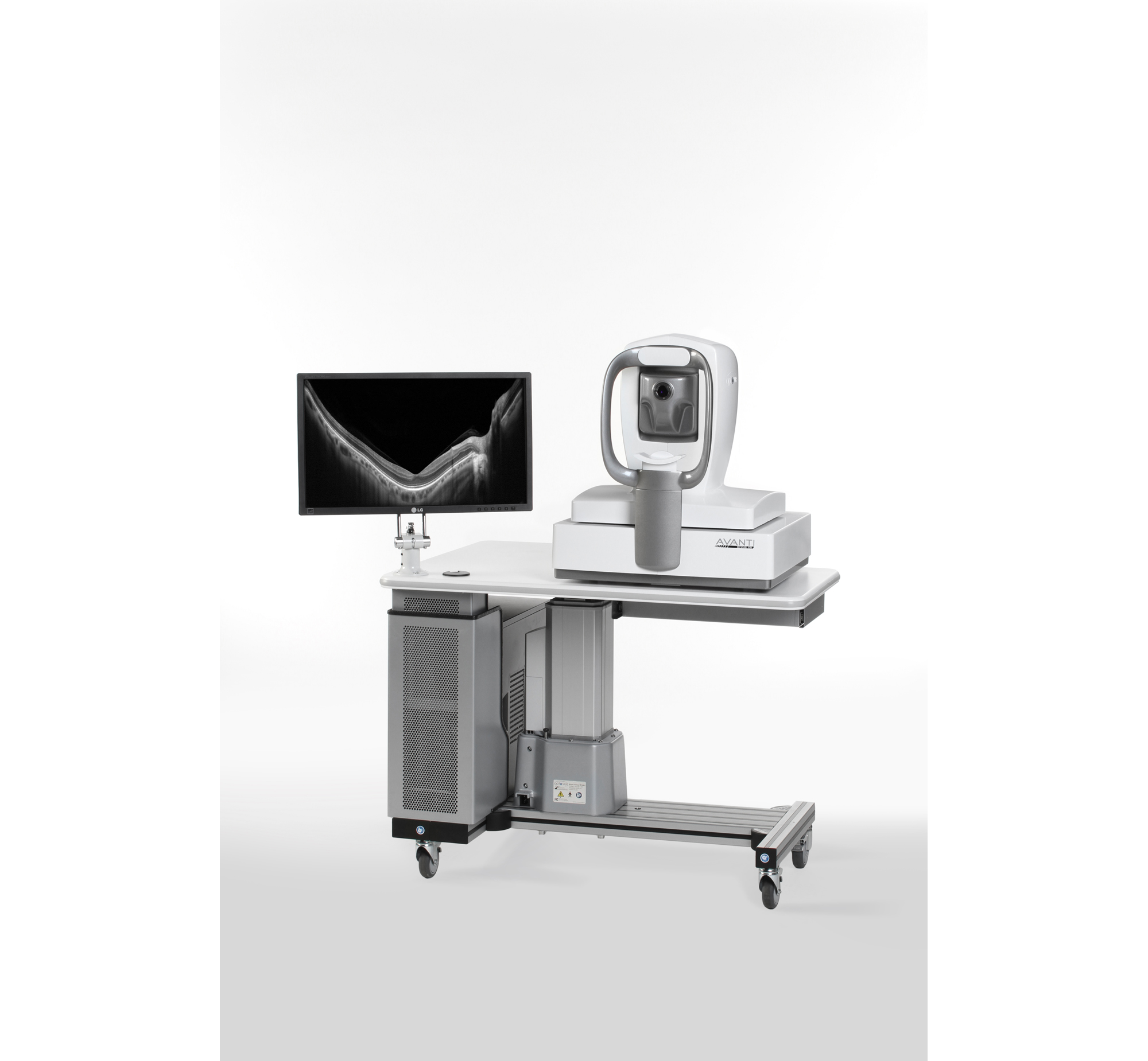
- Assess of change over time with RNFL and GCC trend analysis
- Precisely calculate IOL power in post-refractive surgery patients with Total Cornea Power
- Visualize peripheral pathology can capture over 1,000,000 data points with Avanti's 3D Widefield Cube
- 70,000 A-Scan/Second
- Widefield Scan covers 12 x 9 mm
- Offers non-contact, quantitative measurements of the epithelial and stromal layers of the cornea via epithelial thickness mapping (ETM)
Avanti Widefield OCT provides information on structures outside the traditional 6x6 mm cube, separates the retina into distinct layers for detailed assessment, offers views of the vitreous and deep choroid, and offers the ability to monitor change over time. Cornea Advance enables visualization and measurement of corneal angles and corneal thickness.
No
Not specified
Retina Thickness, Retinal Mapping
—
Normative Data Comparison, Optic Disc, RNFL Analysis, Ganglion Cell Complex
—
Enhanced Depth Imaging (EDI) OCT
—
3D Reconstruction
—
—
—
—
—
- High speed scan
- High resolution image of OCT & SLO
- Imaging Modes: Scanning Laser Imaging
- Imaging Capabilities: Fundus Camera, Wide-Field Imaging, Retina Thickness, Retinal Mapping, RPE Deformation, Corneal Thickness Mapping (optional), Corneal Curvature Mapping (optional)
- Glaucoma Measurements: TSNIT Analysis, Normative Data Comparison, Optic Disc, RNFL Mapping, RNFL Analysis, Ganglion Cell Complex
The RS-3000 Advanced from Nidek delivers high definition allowing the easy detection of subtle fundus structures and pathology. The model includes repeatable follow up scans of reassuring accuracy using Nidek's tracking and vessel recognition technology, and normative database to aid in diagnosis and comparison of images.
Yes
Not specified
Fundus Camera, Wide-Field Imaging, Retina Thickness, Retinal Mapping, RPE Deformation, Corneal Thickness Mapping (Optional), Corneal Curvature Mapping (Optional)
Not specified
TSNIT Analysis, Normative Data Comparison, Optic Disc, RNFL Mapping, RNFL Analysis, Ganglion Cell Complex
Scanning Laser Imaging
Real-Time Video, 3D Reconstruction
Powercord
—
Not specified
—
—

- Powerful diagnostic imaging configuration combines high speed SD-OCT with 5 fundus imaging modes for maximum detection, diagnosis and disease management information
- Upgradable platform with panning camera head
- Delivers precision, detail, and measurement reproducibility of one micron
- Imaging Capabilities: Fundus Camera, Wide-Field Imaging, Retina Thickness, Retinal Mapping, RPE Deformation
- Imaging Modes: Fluorescein Angiography, ICG Angiography, Autofluorescein, Infrared Reflectance, Blue Reflectance, Scanning Laser Imaging
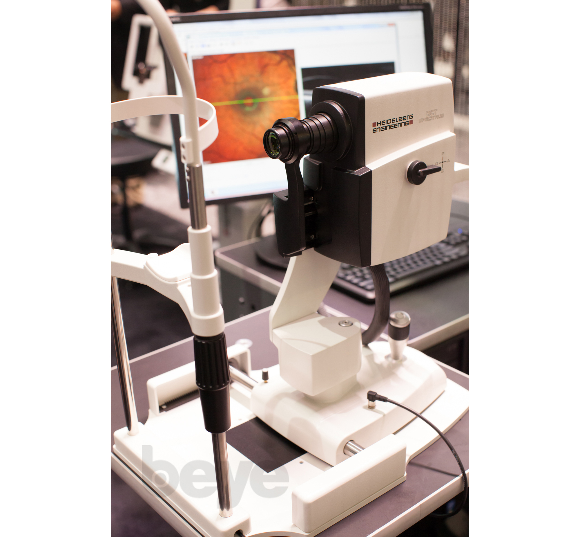
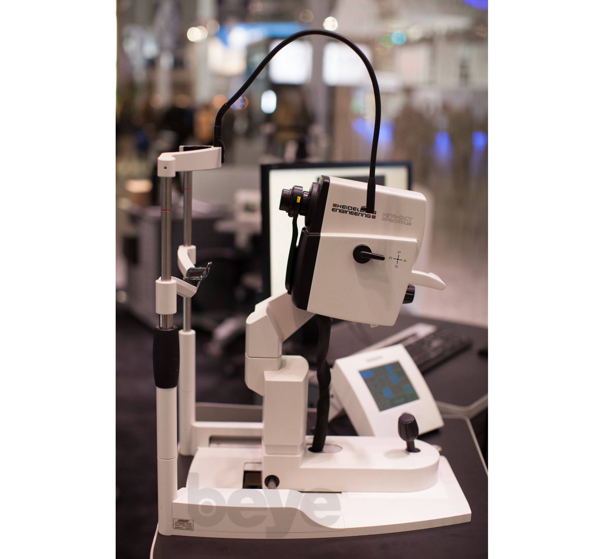
The SPECTRALIS HRA+OCT delivers the ultimate in retinal imaging providing physicians the power of Spectral-domain OCT with TruTrack active eye tracking, AutoRescan features, posterior pole asymmetry analysis combined with five different confocal scanning laser fundus imaging modes and Ultra-Wide Field imaging capability.
Yes
Not specified
Fundus Camera, Wide-Field Imaging, Retina Thickness, Retinal Mapping, RPE Deformation
Not specified
Normative Data Comparison, RNFL Analysis
Fluorescein Angiography, ICG Angiography, Autofluorescence, Infrared Reflectance, Blue Reflectance (Red-Free), Scanning Laser Imaging
Real-Time Video
Powercord
—
Not specified
—
—
- Multi-modality combination of SD-OCT with four fundus imaging modes
- Upgradable platform with panning camera head
- Powered by confocal scanning laser (cSLO) technology for improved image quality, patient comfort, poor dilator effectiveness and clear videos
- Imaging Modes: Fluorescein Angiography, ICG Angiography, Autofluorescein, Infrared Reflectance, Blue Reflectance, Scanning Laser Imaging
- Imaging Capabilities: Fundus Camera, Wide-Field Imaging, Retina Thickness, Retinal Mapping
- Glaucoma Measurements: Normative Data Comparison, RNFL Analysis
The SPECTRALIS FA+OCT provides the diagnostic power of multi-modality imaging by combining Spectral-domain OCT, confocal scanning laser ophthalmoscope (cSLO) fundus imaging, and video angiography.
Yes
Not specified
Fundus Camera, Wide-Field Imaging, Retina Thickness, Retinal Mapping
Not specified
Normative Data Comparison, RNFL Analysis
Fluorescein Angiography, ICG Angiography, Autofluorescence, Infrared Reflectance, Blue Reflectance (Red-Free), Scanning Laser Imaging
Real-Time Video
Powercord
—
Not specified
—
—
- Clear, crisp dynamic angiography
- Upgradable platform with panning camera head to include SD-OCT
- Delivers precision, detail, and versatility
- Imaging Modes: Fluorescein Angiography, ICG Angiography, Autofluorescein, Infrared Reflectance, Blue Reflectance, Scanning Laser Imaging
- Imaging Capabilities: Fundus Camera, Wide-Field Imaging, Retina Thickness,Retinal Mapping
- Glaucoma Measurements: Normative Data Comparison, RNFL Analysis
The multi-modality SPECTRALIS HRA offers both fundus and iris FA as well as simultaneous FA and ICGA. SPECTRALIS video angiography can also be combined with red-free, infrared, and BluePeak blue laser autofluorescence.
Yes
Yes
Fundus Camera, Wide-Field Imaging, Retina Thickness, Retinal Mapping
Not specified
Normative Data Comparison, RNFL Analysis
Fluorescein Angiography, ICG Angiography, Autofluorescence, Infrared Reflectance, Blue Reflectance (Red-Free), Scanning Laser Imaging
Real-Time Video
Powercord
—
Not specified
—
—

- Flexible combination of frequency used in infrared (IR) mode and high speed SD-OCT
- Upgradable Configuration with panning camera head
- Proven technologies deliver precision, detail and measurement reproducibility of one micron
- Normative data comparison
- RNFL analysis
- Imaging Capabilities:
- Fundus camera
- Wide-field imaging
- Retina thickness
- Retinal mapping
The SPECTRALIS OCTPLUS combines Spectral-domain OCT with confocal scanning laser fundus imaging. Like all SPECTRALIS models, the OCTPLUS includes TruTrack active eye tracking and AutoRescan features.
Yes
Yes
Fundus Camera, Wide-Field Imaging, Retina Thickness, Retinal Mapping
Not specified
Normative Data Comparison, RNFL Analysis
Infrared Reflectance, Scanning Laser Imaging
Not specified
Powercord
—
Not specified
—
—

- Flexible Workflow Integration
- Multi-modality platform
- 3D Volume scans with complete raster averaging
- Imaging Modes: Fluorescein Angiography, ICG Angiography, Autofluorescein, Infrared Reflectance, Blue Reflectance, Scanning Laser Imaging
- Imaging Capabilities: Fundus Camera, Wide-Field Imaging, Retina Thickness, Retinal Mapping, RPE Deformation
- Glaucoma Measurements: Normative Data Comparison, Optic Disc, RNFL Mapping, RNFL Analysis
- 3D Reconstruction
The HRA+OCT is a non-contact ophthalmic diagnostic imaging device. It is intended for viewing the posterior segment of the eye, including: two and three dimensional imaging, cross sectional imaging, fundus photography, and fluorescence imaging.
Yes
Yes
Fundus Camera, Wide-Field Imaging, Retina Thickness, Retinal Mapping, RPE Deformation
Not specified
Normative Data Comparison, Optic Disc, RNFL Mapping, RNFL Analysis
Enhanced Depth Imaging (EDI) OCT, Fluorescein Angiography, Infrared Reflectance, Blue Reflectance (Red-Free), Scanning Laser Imaging
3D Reconstruction
Powercord
—
Not specified
—
—
- Single-shot fundus autofluorescence
- Great detail density
- Interactive review
- Multimodal assessments
- Camera Modes: Fluorescein Aniography, Autofluorescence, Infrared Reflectance, Blue Reflectance (red-free), Scanning Laser Imaging
- Imaging Capabilities: Fundus camera, Retina thickness, Corneal thickness mapping
- Size is 16.1 (W) x 18.9 (D) x 26.8 (H) in. and weight is 32.9 kg (72.7 lbs.)
CIRRUS photo combines a mydriatic/non-mydriatic fundus camera with CIRRUS HD-OCT technology in one compact and versatile system. Available in two models, CIRRUS photo 600 and CIRRUS photo 800, it provides multiple insights for comprehensive retina and posterior segment care.
Yes
Yes
Fundus Camera, Retina Thickness, Corneal Thickness Mapping
—
Normative Data Comparison, RNFL Analysis
—
Fluorescein Angiography, ICG Angiography, Autofluorescence, Infrared Reflectance, Blue Reflectance (Red-Free), Scanning Laser Imaging
—
Not specified
—
Standalone
—
Mydriatic
—
—
—
Privacy | Terms & Conditions Ⓒ 2026 Beye, LLC. All rights reserved.
