- Product Listings
- Compare Products
- Calculators
- Product Tours
- Expert Reviews
- Our Experts
- Company Directory
- About Beye
- Contact Us
Ⓒ 2026 Beye.com. All rights reserved.
This content is intended for health care professionals and providers only. The information contained on Beye.com, including text, graphics, images, and interactive activities, is for informational purposes only, and is not intended to be a substitute for professional medical advice. Beye LLC, via its Editors and Publisher, accepts no responsibility for any injury or damage to persons or property occasioned through the implementation of any ideas or use of any product described herein. Although great care is taken to ensure that all information is accurate, it is recommended that readers seek independent verification of advice on drugs and other product usage, surgical techniques and clinical processes prior to their use. References made in article may indicate usage of medical equipment or drugs at dosages, for periods of time, and in combination not included in the current prescribing information. Inclusion of advertising materials on the website thereof, does not constitute and representation or guarantee by Beye LLC of the quality of such products, or of the claims made.
Compare Intraoperative OCTs
3 Products
reset all
At-a-Glance
Description
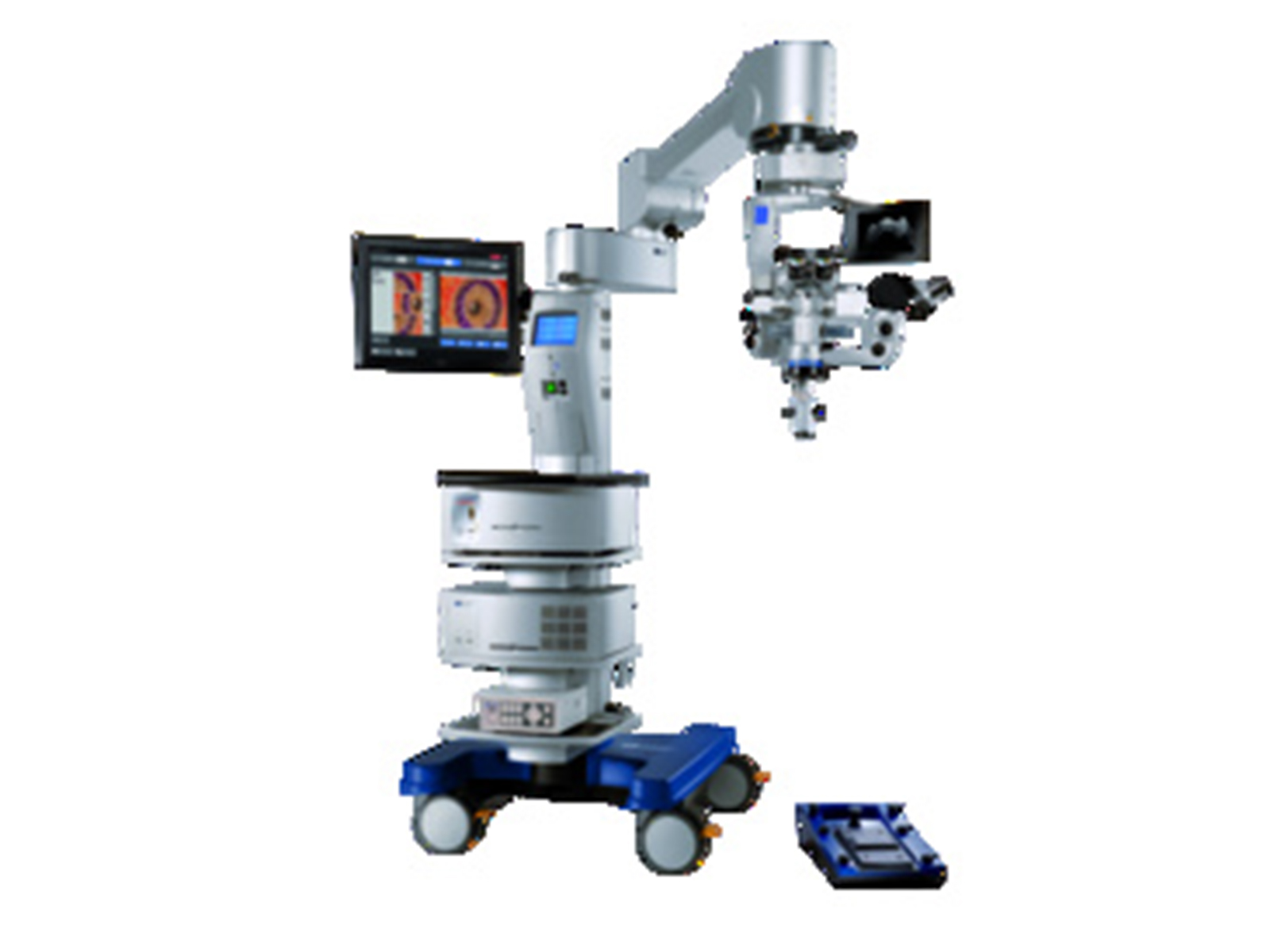
- Can select between Xenon Superlux eye, LED, and established halogen light sources
- Integrated slit illuminator
- RESIGHT 700 fundus viewing system allows surgeon to recognize details of the retina
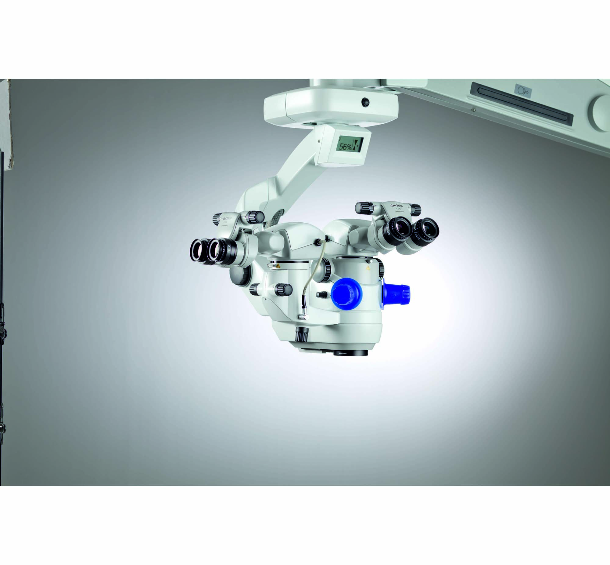
- Binocular Tube: Apochromatic optics, invertertube
- Eyepiece: 10x
- Magnification Changes:Electric/motorized, focus range: 70 mm
- Total Magnification: 0.4 - 2.4x
- Blue blocking (all light sources), HaMode (Superlux Eye), HaMode (LED fiber optic), and 25% gray (LED fiber optic) filters
Used for cataract and retina procedures, the OPMI LUMERA 700 and RESCAN 700 has a wide range of customization options. The microscope utilizes the Stereo Coaxial Illumination (SCI) system for red reflex and image quality.
HAAG-STREIT SURGICAL introduced the world‘s first intraoperative OCT system, developed by OPMedT. OCT scans of the eye's anterior and posterior segments are performed live during surgery, thus increasing the patient's safety and improving quality control mechanisms for the surgeon.
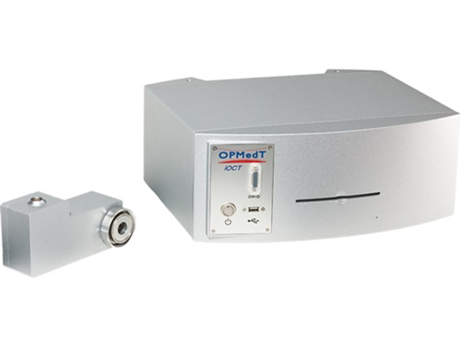
The Optical Coherence Tomography (OCT) is a new medical imaging device. It uses ultrasound with light and enables user to visualize small structures and layers inside the tissue with a resolution of about 10 µm.
Privacy | Terms & Conditions Ⓒ 2026 Beye, LLC. All rights reserved.
