- Product Listings
- Compare Products
- Calculators
- Product Tours
- Expert Reviews
- Our Experts
- Company Directory
- About Beye
- Contact Us
Ⓒ 2026 Beye.com. All rights reserved.
This content is intended for health care professionals and providers only. The information contained on Beye.com, including text, graphics, images, and interactive activities, is for informational purposes only, and is not intended to be a substitute for professional medical advice. Beye LLC, via its Editors and Publisher, accepts no responsibility for any injury or damage to persons or property occasioned through the implementation of any ideas or use of any product described herein. Although great care is taken to ensure that all information is accurate, it is recommended that readers seek independent verification of advice on drugs and other product usage, surgical techniques and clinical processes prior to their use. References made in article may indicate usage of medical equipment or drugs at dosages, for periods of time, and in combination not included in the current prescribing information. Inclusion of advertising materials on the website thereof, does not constitute and representation or guarantee by Beye LLC of the quality of such products, or of the claims made.
Compare Specular Microscopes
8 Products
reset all
At-a-Glance
Description
FDA
CE Mark
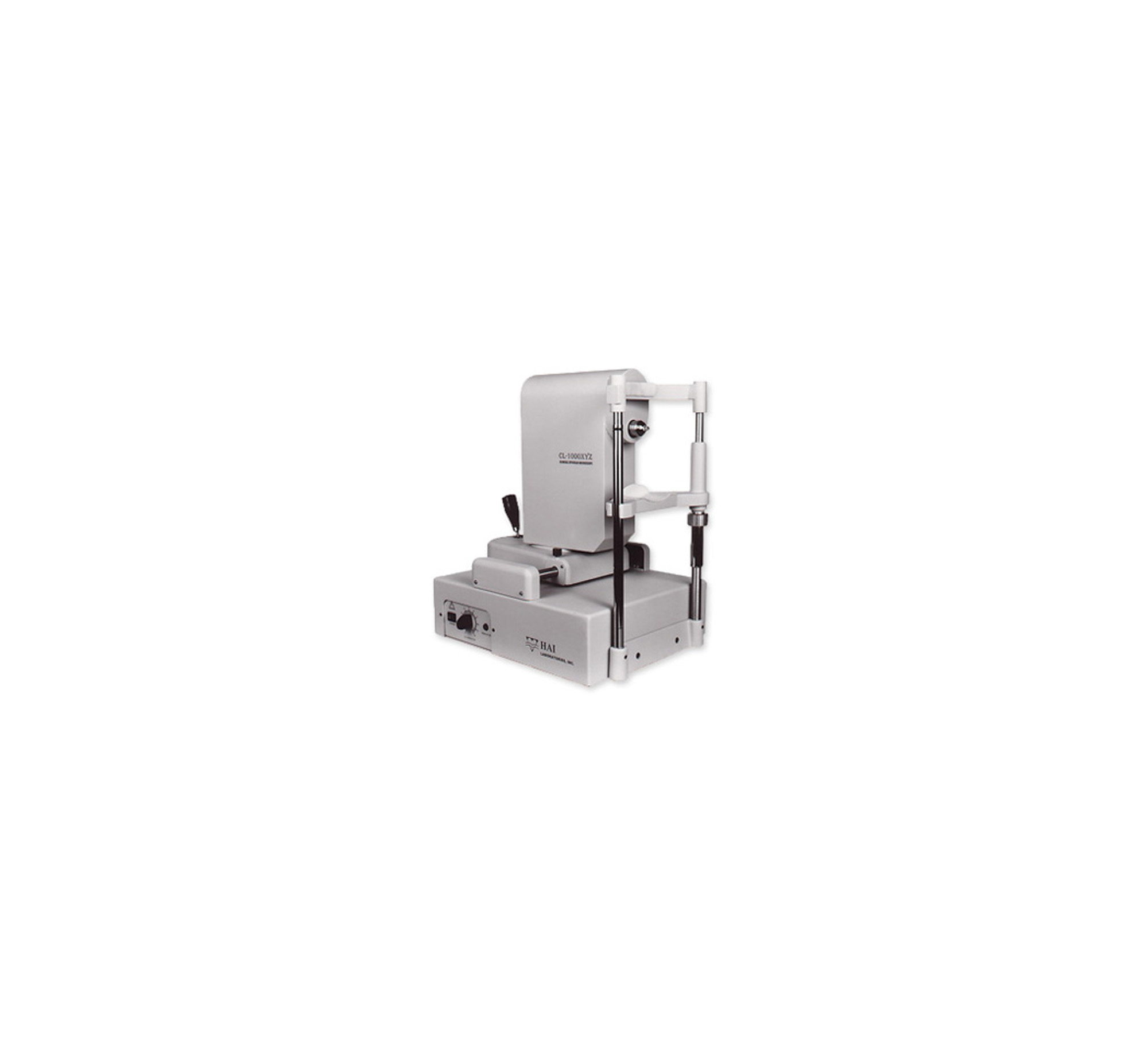
- Rapid image acquisition
- Optional digital pachymeter
- Observation from epithelium to endothelium
- Low-level illumination for patient comfort
- Photographic Coverage is 250µm x 400µm
- 30 images per pass
The HAI CL-1000xyz Contact Clinical Specular Microscope's focal depth of 0-999µm gives a clear image no matter what disease or cornea thickness, making this scope ideal for post-DSAEK follow-up and for patients with edema, severe dystrophy or other abnormal conditions.
Yes
Not specified
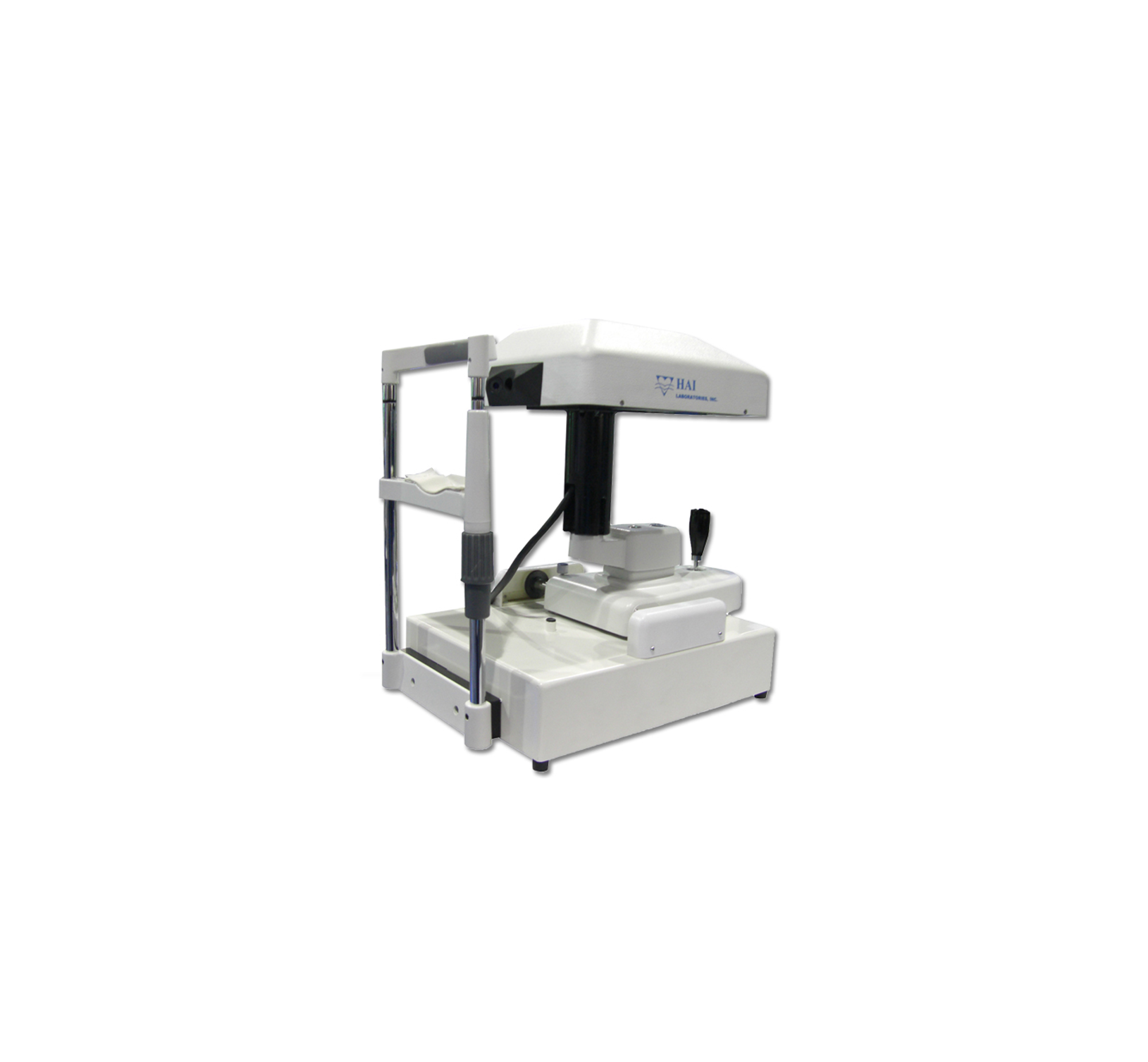
- Live image of endothelium and digital image and data archival
- Auto selection of good endothelial images
- Center and peripheral measuring points
- Photographic Coverage is 300µm x 600µm
- Measurements: Analysis (Auto-count, Manual adjust)
The CL-1000nc is quick and easy to use, capturing endothelial cell images automatically without needing to click any buttons during target acquisition. The software provides quick reference data for cell density, coefficient of variation and hexagonal percentage within a matter of seconds, while a built-in pachymeter gives the thickness of the cornea.
Yes
Yes
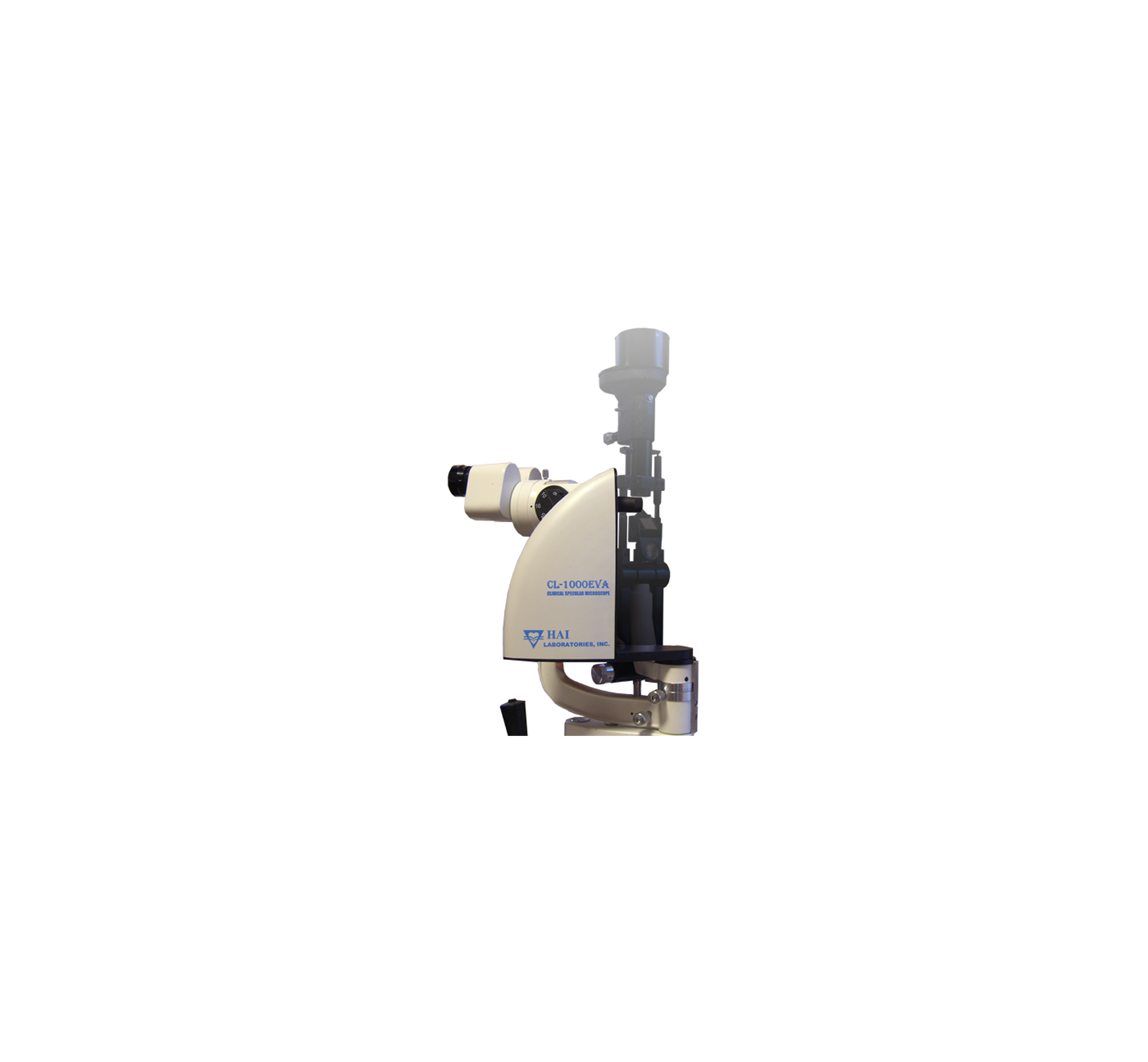
- Live image of endothelium
- Auto selection of good endothelial images
- Analysis includes up to 200 cells
- Cell density, cell area and morphology
- Measurements: Endothelial Cell Density, Endothelial Cell Count
The HAI CL-1000eva Endothelium Viewing Attachment converts your slit lamp into a specular microscope, providing a high-magnification view of endothelial cells with software that performs automatic image capture, frame selection, enhancement and analysis.
Yes
Yes
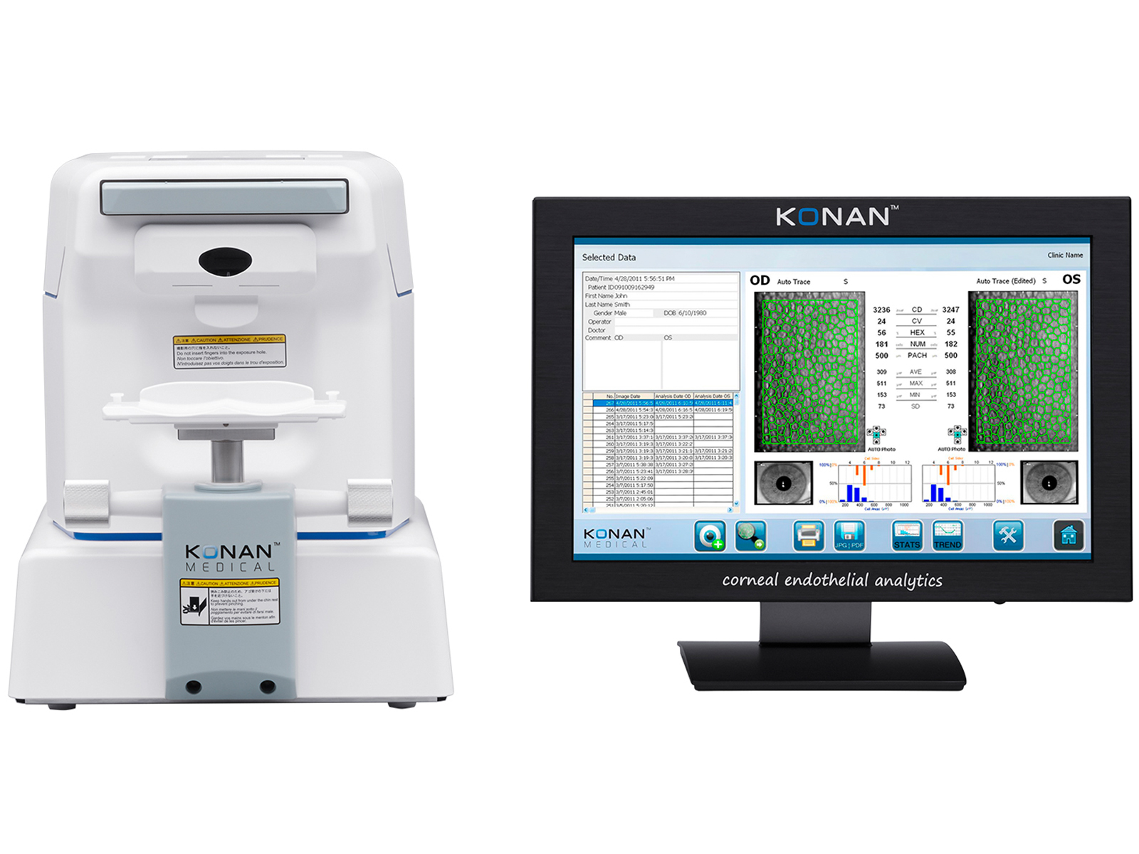
- Flash type is Konan Xe tube
- Photographic Coverage is 0.1mm squared
- Measurements: Endothelial Cell Density, Corneal Thickness, Corneal Thickness Measurements Accuracy (±10m or better), Endothelial Cell Count, Analysis (Auto-analysis)
- Built-in color printer
While simple to use, the CellChek XL provides practitioners with critical information for procedures including corneal tissue transplantations, refractive IOLs, contact lens fitting and many others. The testing, from imaging to automated analysis, fits easily into your patient flow.
Yes
Yes
- Paracentral specular microscopy
- 3-D auto tracking and auto shot
- 16 images per pass
- Photographic Coverage is 0.25mm x 0.55mm
- Measurements: Endothelial Cell Density, Analysis (Two-second Auto-analysis)
- Instant printout with built-in printer
The CEM-530 includes a unique function to capture paracentral images. The paracentral images are captured at eight points, 5° visual angle within a 0.25 mm x 0.55 mm field and enable enhanced assessment surrounding the central image.
Yes
Yes
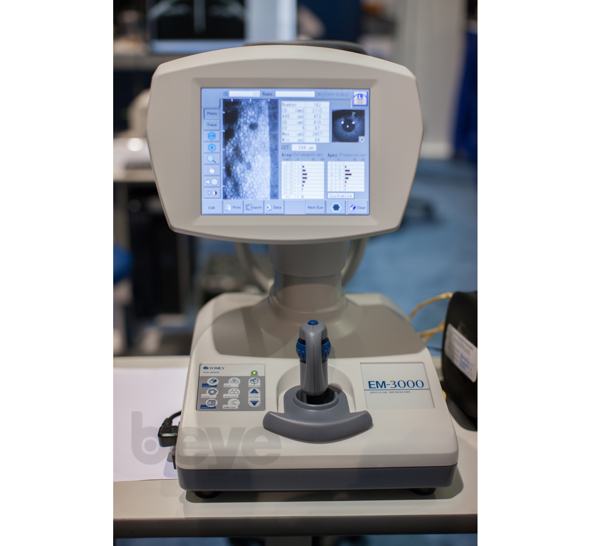
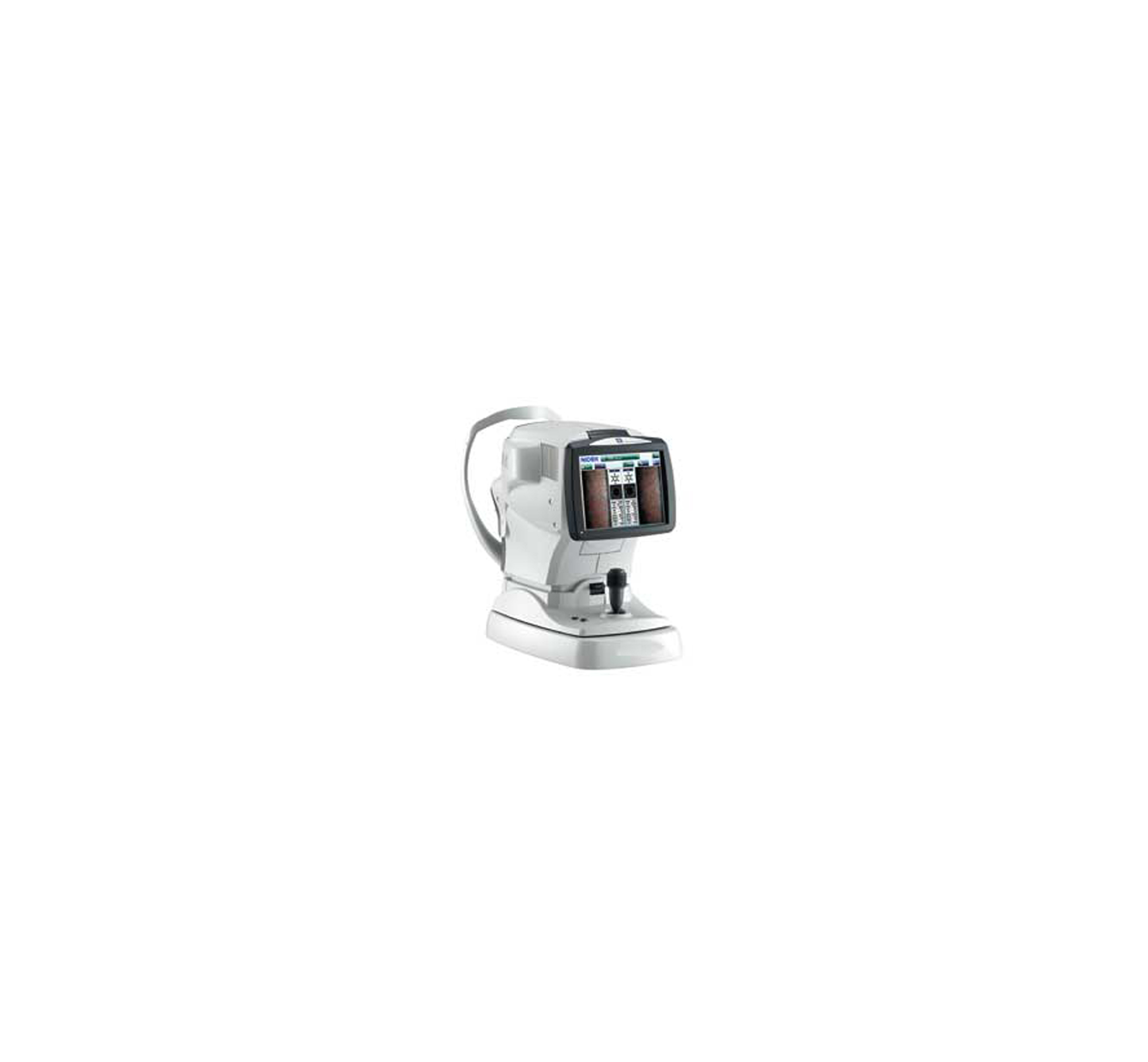
- Easy photographing using the touch alignment
- Serial photographs of 15 shots with 7 capturing positions
- Flash type is LED light source
- Photographic Coverage is 0.25mm x 0.54mm
- Measurements: Endothelial Cell Density, Corneal Thickness, Corneal Thickness Measurement Accuracy (±10 µm), Endothelial Cell Count, Analysis
- Built-in printer
Tomey USA's EM 3000 specular microscope combines a fully automated color touch screen feature with an easy-to-use client interface that offers superior imaging of the corneal region. The EM 3000 provides ease of operation with its performance accuracy with the incorporation of auto alignment and auto shot functions at the touch of a finger.
Yes
Not specified
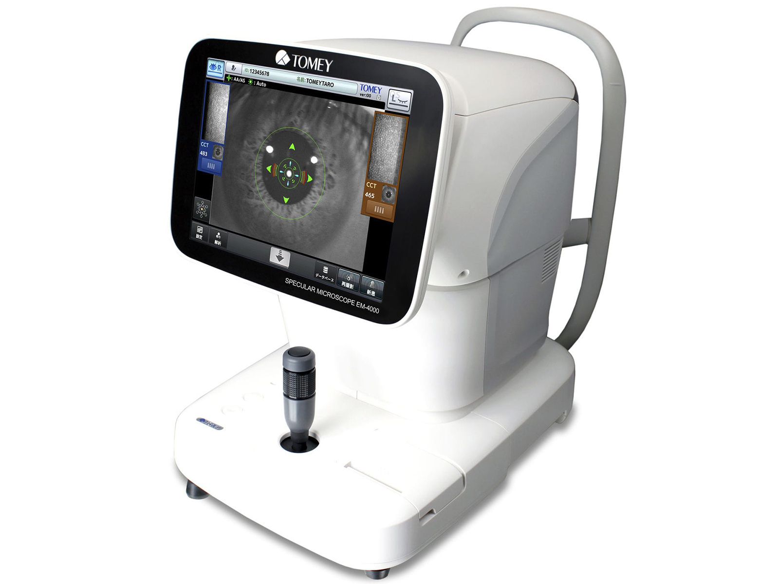
- Auto elignment + auto measurement and integrated non-contact Pachymetry
- 13 measurement areas
- Measurements: Endothelial Cell Density, Corneal Thickness, Endothelial Cell Count (Up to 300), Analysis (CD (cell density), AVG (average cell area), SD (standard deviation of cell area), CV (coefficient of variation of cell area), Cell size (max. + min. cell area))
- Built-in printer
The Specular Microscope EM-4000 provides corneal endothelium images by using the specular optical principle with full automatic alignment. EM-4000 uses LED light source and a high-speed CCD camera to capture 16 images in series. It automatically selects and instantly displays the finest one from these images. It can perform automatic analysis as well as two manual analyses "Core method" and "L count method" with built-in software.
Not specified
Not specified
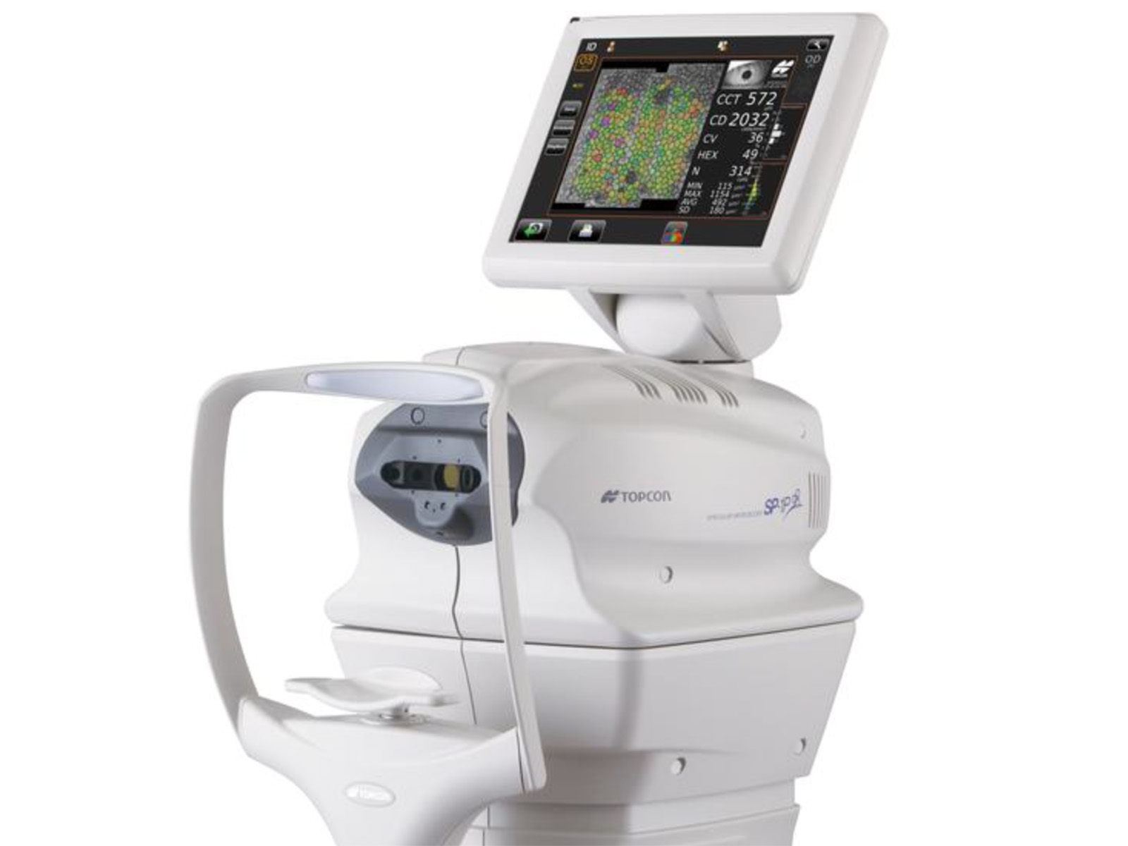
- Wide angle "panorama" photography mode
- Two specific photography modes - sequence course & free style course
- Photographic Coverage is 0.25x0.55mm
- Measurements: Corneal Thickness, Corneal Thickness Measurement Accuracy (Measured only when taking a photograph using the central fixation target)
The new Topcon Specular Microscope model SP-1P* introduces a fully automatic capture procedure together with a modern, ergonomic design that simplifies its use and increases its efficiency. A large 10.4 inch rotatable touch panel monitor eliminates the need for a joystick.
Yes
Yes
Privacy | Terms & Conditions Ⓒ 2026 Beye, LLC. All rights reserved.
