- Product Listings
- Compare Products
- Calculators
- Product Tours
- Expert Reviews
- Our Experts
- Company Directory
- About Beye
- Contact Us
Ⓒ 2026 Beye.com. All rights reserved.
This content is intended for health care professionals and providers only. The information contained on Beye.com, including text, graphics, images, and interactive activities, is for informational purposes only, and is not intended to be a substitute for professional medical advice. Beye LLC, via its Editors and Publisher, accepts no responsibility for any injury or damage to persons or property occasioned through the implementation of any ideas or use of any product described herein. Although great care is taken to ensure that all information is accurate, it is recommended that readers seek independent verification of advice on drugs and other product usage, surgical techniques and clinical processes prior to their use. References made in article may indicate usage of medical equipment or drugs at dosages, for periods of time, and in combination not included in the current prescribing information. Inclusion of advertising materials on the website thereof, does not constitute and representation or guarantee by Beye LLC of the quality of such products, or of the claims made.
Compare Swept-Source OCTs
5 Products
reset all
At-a-Glance
Description
FDA
CE Mark
Capabilities
Measurements
Modes
Processing
- Visualize vessels of the retina and choroid non-invasively
- Analyze structure and function with a single imaging platform
- Personalize your approach to each patient by imaging as often as needed
AngioVue Retina is a unique platform designed specifically for retina applications that combines functional OCT Angiography (OCTA) with the structural OCT retina scans your practice demands. AngioVue Retina integrates OCTA, without replacing your existing structural OCT system, in a flexible and modular approach. In addition, you'll expand your imaging capacity to prevent workflow bottlenecks in the pre-test area.
Yes
Yes
Wide-Field Imaging, Retina Thickness, Retinal Mapping, RPE Deformation, Corneal Thickness Mapping, Corneal Curvature Mapping
Normative Data Comparison, Optic Disc, Ganglion Cell Complex
Enhanced Depth Imaging (EDI) OCT, Fluorescein Angiography, Autofluorescence, Scanning Laser Imaging
Depth-resolved imaging displays individual layers of the retina

- Visualize vessels of the retina and choroid non-invasively
- AngioVueHD 6x6 mm scan or assessment over a wider field of view
AngioVueHD provides a 6x6 mm scan with outstanding resolution to evaluate fine vessels over a larger section of the retina. Automatic montage instantly combines macula and optic disc images for high-density widefield visualization (10x6 mm).
Yes
No
Not specified
Not specified
Not specified
Not specified
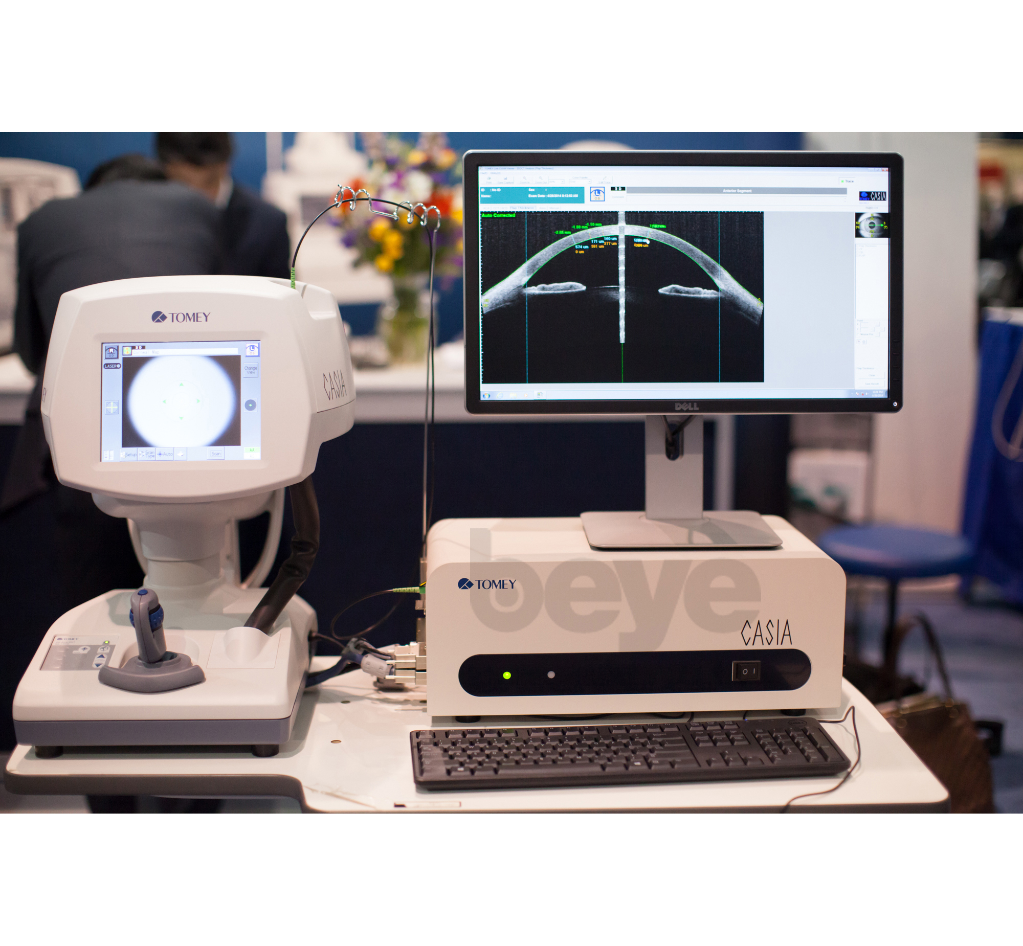
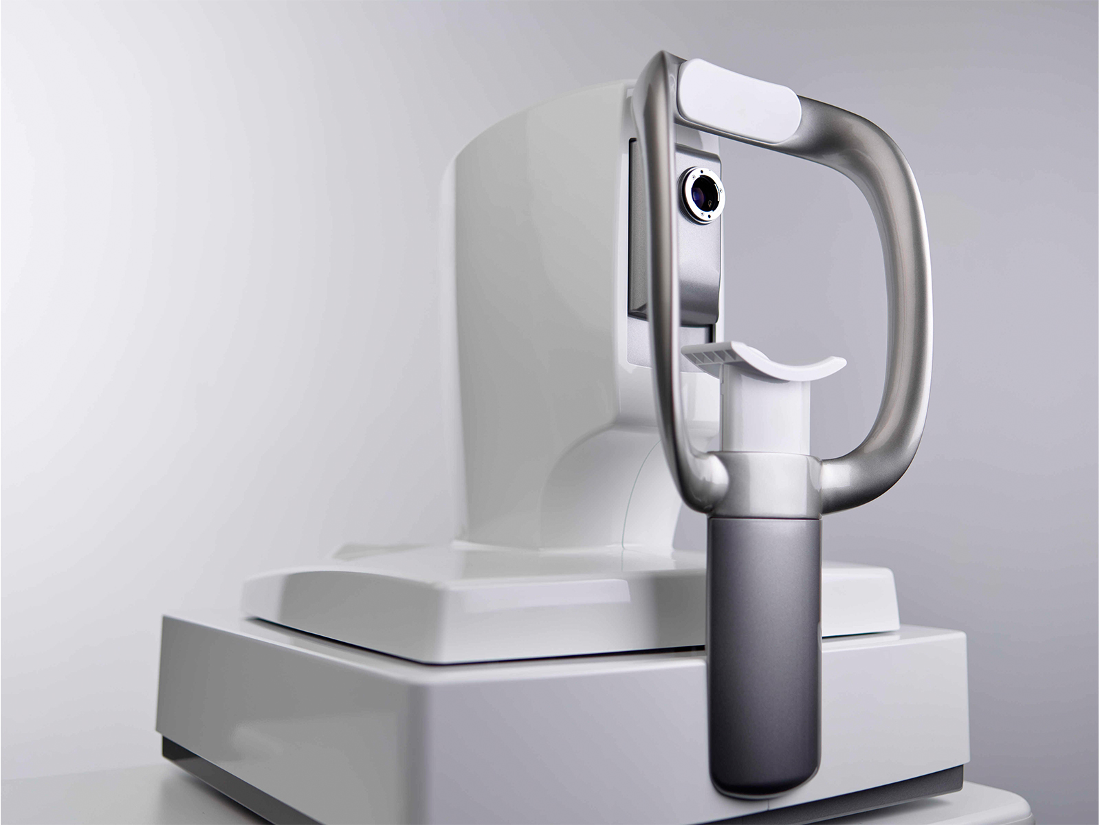
- High resolution images of the arbitrary area at high speed
- Less motion artifacts with fast acquisition
- Fulfillment of the useful application and utility software
- Auto alignment / Auto shot
- Corneal shape analysis
- 17 inch color display
The CASIA Corneal/Anterior Segment OCT SS-1000 is a non-contact, non-invasive three dimensional imaging swept source OCT system. This system achieves high resolution and high speed. The CASIA is indicated for cross sectional imaging of the anterior segment components of the human eye, and also for dimension measurements such as curvature, length, area and volume by computed analysis.
No
Not specified
Corneal Thickness Mapping, Corneal Curvature Mapping
Not specified
Not specified
3D Reconstruction (Optional), 2D mode, Movie mode, Custom scan
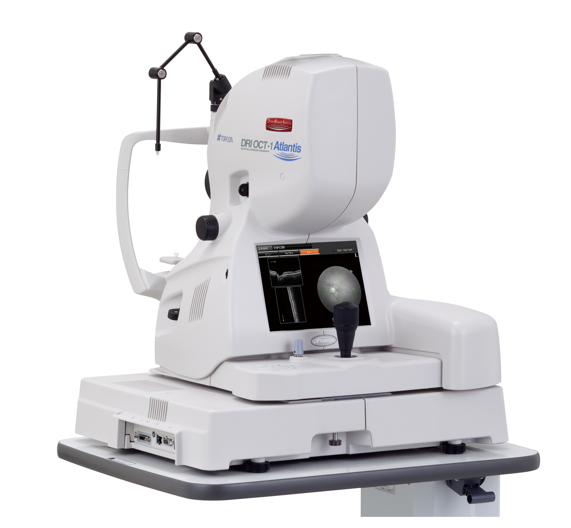
- 1,050 nm wavelength provides deep range imaging
- 100,000 A-Scans/sec - unsurpassed scanning speed
- Invisible scan lines
- 12 mm wide Scan
- 1 micron wavelength for deeper penetration into choroid and sclera with less light scattering and uniform sensitivity
Topcon has developed DRI OCT-1, a swept source OCT for posterior imaging, utilizing a wavelength of 1,050 nm. It has a fast scanning speed of 100,000 A-scans/sec. Utilizing this 1,050 nm wavelength, DRI OCT-1 can penetrate deeper compared to the current conventional OCT's with wavelength in the 850 nm range. The DRI OCT-1 is meant for Deep Range Imaging for research purposes.
No
Yes
Fundus Camera, Retina Thickness, Retinal Mapping
RNFL Mapping, RNFL Analysis
Infrared Reflectance, Blue Reflectance (Red-Free)
3D Reconstruction (Optional)
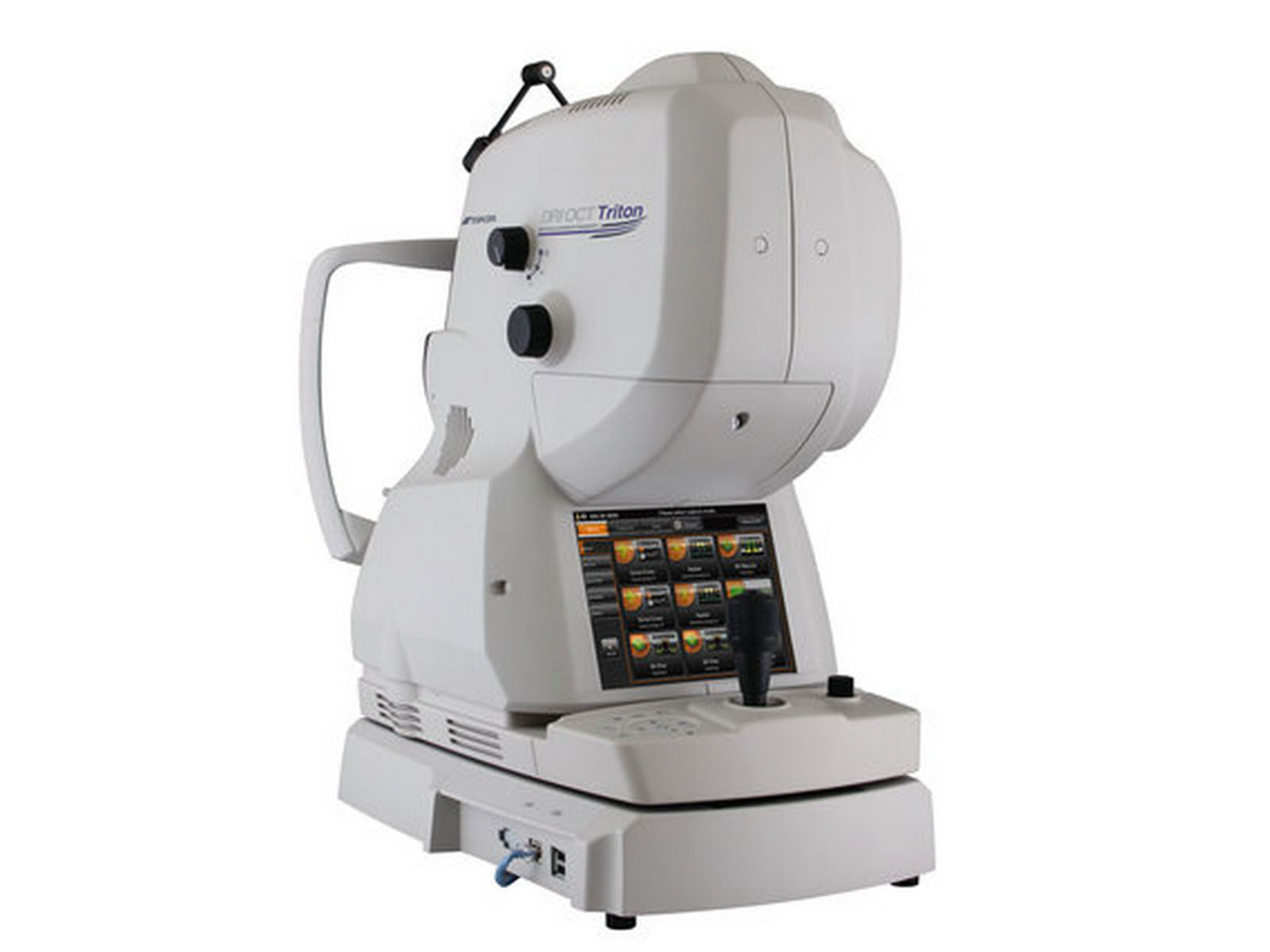
- Single scan capture of comprehensive data
The DRI OCT Triton was the first combined anterior and posterior swept source OCT. The DRI OCT Triton incorporates full color high-resolution fundus photography and FA & FAF imaging. FA & FAF imaging is a factory option.
Not specified
Yes
Fundus Camera
Not specified
Fluorescein Angiography, Infrared Reflectance, Blue Reflectance (Red-Free)
Not specified
Privacy | Terms & Conditions Ⓒ 2026 Beye, LLC. All rights reserved.
