- Product Listings
- Compare Products
- Calculators
- Product Tours
- Expert Reviews
- Our Experts
- Company Directory
- About Beye
- Contact Us
Ⓒ 2026 Beye.com. All rights reserved.
This content is intended for health care professionals and providers only. The information contained on Beye.com, including text, graphics, images, and interactive activities, is for informational purposes only, and is not intended to be a substitute for professional medical advice. Beye LLC, via its Editors and Publisher, accepts no responsibility for any injury or damage to persons or property occasioned through the implementation of any ideas or use of any product described herein. Although great care is taken to ensure that all information is accurate, it is recommended that readers seek independent verification of advice on drugs and other product usage, surgical techniques and clinical processes prior to their use. References made in article may indicate usage of medical equipment or drugs at dosages, for periods of time, and in combination not included in the current prescribing information. Inclusion of advertising materials on the website thereof, does not constitute and representation or guarantee by Beye LLC of the quality of such products, or of the claims made.
Compare Scanning Laser Ophthalmoscope
13 Products
reset all
At-a-Glance
Description
FDA
CE Mark
Capabilities
Image Capture
Measurements
Modes
Processing
Source
System
Video Capture

- Flexible Workflow Integration
- Multi-modality platform
- 3D Volume scans with complete raster averaging
- Imaging Modes: Fluorescein Angiography, ICG Angiography, Autofluorescein, Infrared Reflectance, Blue Reflectance, Scanning Laser Imaging
- Imaging Capabilities: Fundus Camera, Wide-Field Imaging, Retina Thickness, Retinal Mapping, RPE Deformation
- Glaucoma Measurements: Normative Data Comparison, Optic Disc, RNFL Mapping, RNFL Analysis
- 3D Reconstruction
The HRA+OCT is a non-contact ophthalmic diagnostic imaging device. It is intended for viewing the posterior segment of the eye, including: two and three dimensional imaging, cross sectional imaging, fundus photography, and fluorescence imaging.
Yes
Yes
Fundus Camera, Wide-Field Imaging, Retina Thickness, Retinal Mapping, RPE Deformation
Not specified
Normative Data Comparison, Optic Disc, RNFL Mapping, RNFL Analysis
Enhanced Depth Imaging (EDI) OCT, Fluorescein Angiography, Infrared Reflectance, Blue Reflectance (Red-Free), Scanning Laser Imaging
3D Reconstruction
Powercord
—
Not specified
—

- Flexible combination of frequency used in infrared (IR) mode and high speed SD-OCT
- Upgradable Configuration with panning camera head
- Proven technologies deliver precision, detail and measurement reproducibility of one micron
- Normative data comparison
- RNFL analysis
- Imaging Capabilities:
- Fundus camera
- Wide-field imaging
- Retina thickness
- Retinal mapping
The SPECTRALIS OCTPLUS combines Spectral-domain OCT with confocal scanning laser fundus imaging. Like all SPECTRALIS models, the OCTPLUS includes TruTrack active eye tracking and AutoRescan features.
Yes
Yes
Fundus Camera, Wide-Field Imaging, Retina Thickness, Retinal Mapping
Not specified
Normative Data Comparison, RNFL Analysis
Infrared Reflectance, Scanning Laser Imaging
Not specified
Powercord
—
Not specified
—
- Clear, crisp dynamic angiography
- Upgradable platform with panning camera head to include SD-OCT
- Delivers precision, detail, and versatility
- Imaging Modes: Fluorescein Angiography, ICG Angiography, Autofluorescein, Infrared Reflectance, Blue Reflectance, Scanning Laser Imaging
- Imaging Capabilities: Fundus Camera, Wide-Field Imaging, Retina Thickness,Retinal Mapping
- Glaucoma Measurements: Normative Data Comparison, RNFL Analysis
The multi-modality SPECTRALIS HRA offers both fundus and iris FA as well as simultaneous FA and ICGA. SPECTRALIS video angiography can also be combined with red-free, infrared, and BluePeak blue laser autofluorescence.
Yes
Yes
Fundus Camera, Wide-Field Imaging, Retina Thickness, Retinal Mapping
Not specified
Normative Data Comparison, RNFL Analysis
Fluorescein Angiography, ICG Angiography, Autofluorescence, Infrared Reflectance, Blue Reflectance (Red-Free), Scanning Laser Imaging
Real-Time Video
Powercord
—
Not specified
—
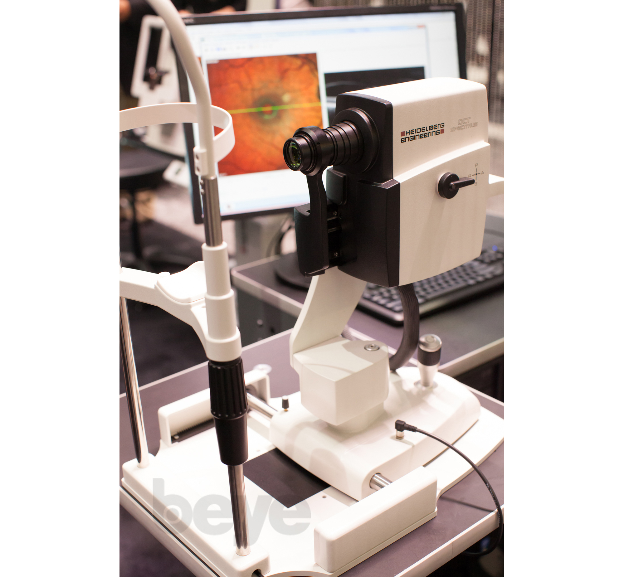
- Multi-modality combination of SD-OCT with four fundus imaging modes
- Upgradable platform with panning camera head
- Powered by confocal scanning laser (cSLO) technology for improved image quality, patient comfort, poor dilator effectiveness and clear videos
- Imaging Modes: Fluorescein Angiography, ICG Angiography, Autofluorescein, Infrared Reflectance, Blue Reflectance, Scanning Laser Imaging
- Imaging Capabilities: Fundus Camera, Wide-Field Imaging, Retina Thickness, Retinal Mapping
- Glaucoma Measurements: Normative Data Comparison, RNFL Analysis
The SPECTRALIS FA+OCT provides the diagnostic power of multi-modality imaging by combining Spectral-domain OCT, confocal scanning laser ophthalmoscope (cSLO) fundus imaging, and video angiography.
Yes
Not specified
Fundus Camera, Wide-Field Imaging, Retina Thickness, Retinal Mapping
Not specified
Normative Data Comparison, RNFL Analysis
Fluorescein Angiography, ICG Angiography, Autofluorescence, Infrared Reflectance, Blue Reflectance (Red-Free), Scanning Laser Imaging
Real-Time Video
Powercord
—
Not specified
—

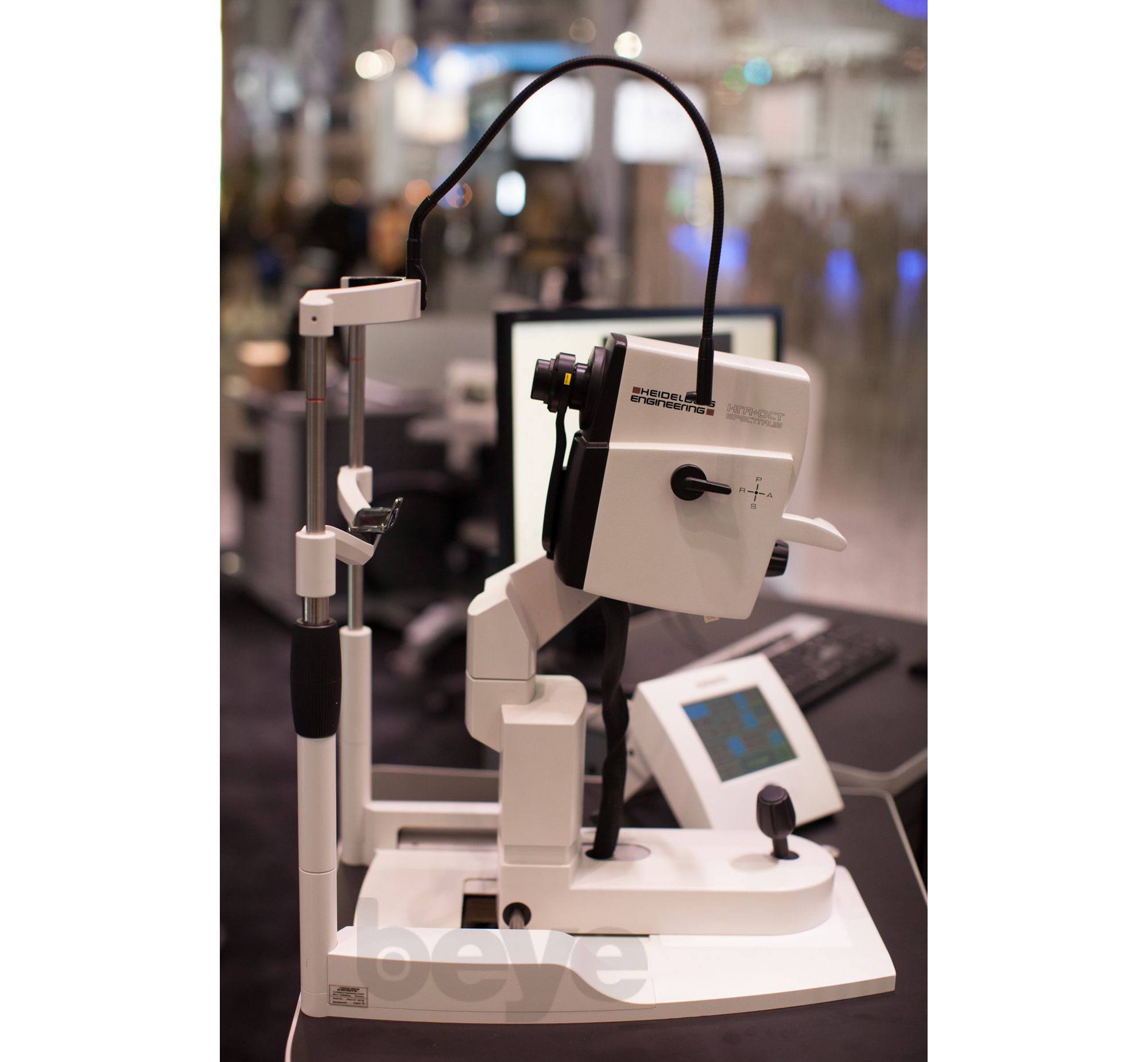
- Powerful diagnostic imaging configuration combines high speed SD-OCT with 5 fundus imaging modes for maximum detection, diagnosis and disease management information
- Upgradable platform with panning camera head
- Delivers precision, detail, and measurement reproducibility of one micron
- Imaging Capabilities: Fundus Camera, Wide-Field Imaging, Retina Thickness, Retinal Mapping, RPE Deformation
- Imaging Modes: Fluorescein Angiography, ICG Angiography, Autofluorescein, Infrared Reflectance, Blue Reflectance, Scanning Laser Imaging
The SPECTRALIS HRA+OCT delivers the ultimate in retinal imaging providing physicians the power of Spectral-domain OCT with TruTrack active eye tracking, AutoRescan features, posterior pole asymmetry analysis combined with five different confocal scanning laser fundus imaging modes and Ultra-Wide Field imaging capability.
Yes
Not specified
Fundus Camera, Wide-Field Imaging, Retina Thickness, Retinal Mapping, RPE Deformation
Not specified
Normative Data Comparison, RNFL Analysis
Fluorescein Angiography, ICG Angiography, Autofluorescence, Infrared Reflectance, Blue Reflectance (Red-Free), Scanning Laser Imaging
Real-Time Video
Powercord
—
Not specified
—
- Multiple applications: glaucoma, retinal edema, cornea/conjunctiva
- Laptop or PC based
- Field of view is 15° x 15° (transversal)
- Focus range is -12 to +12 dpt. (sphere), -6 to +6 dpt. (cylinder; astigmatism lenses)
- Optical resolution is approx. 10 μm (transversal) x 300 μm (longitudinal)
- EMR compatible
- Pupil diameter is >= 1 mm
- Frame rate is 1–6 seconds per 3-D image
- Light source: internal and external fixation lamp
HRT3 is a confocal laser scanning system, designed to capture and analyze three-dimensional images of the posterior segment of the eye. It allows a quantitative assessment of the topography in the structures of the eye, especially of the retinal topography, as well as the precise tracking of any topographical changes. It is applied more often to describe the glaucomatous optic nerve head, the analysis of the slits and the edema of the macula and the analysis in the layer of the nerve fibers.
Not specified
Not specified
—
Yes
—
Powercord
—
No
—
—
—
—
—

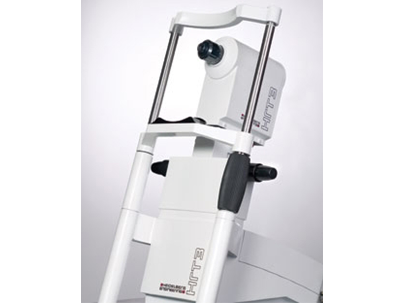
- Laptop or PC based
- Field of view is 15° x 15° (transversal)
- Focus range is -12 to +12 dpt. (sphere), -6 to +6 dpt. (cylinder; astigmatism lenses)
- Optical resolution is approx. 10 μm (transversal) x 300 μm (longitudinal)
- EMR compatible
- Pupil diameter is >= 1 mm
- Light source: internal and external fixation lamp
The Heidelberg Retina Tomograph (HRT) is a tool for detecting and managing glaucoma, especially for assisting in the identification of pre-perimetric disease and tracking progression. The Ancillary Study to the Ocular Hypertension Treatment Study (OHTS) demonstrated that optic disc analysis detected glaucoma conversion in 55% of cases, before any detectable loss of visual function. The HRT has proved to be a robust predictor of glaucoma.
Not specified
Not specified
—
Yes
—
Powercord
—
No
—
—
—
—
—

- Retinal thickness mapping in 3D
- Precise detection of change with TruTrack image alignment
- No interpolated data, less chance of missing pathology
- Field of view is 15° x 15° (transversal)
- Focus range is -12 to +12 dpt. (sphere), -6 to +6 dpt. (cylinder; astigmatism lenses)
- Optical resolution is approx. 10 μm (transversal) x 300 μm (longitudinal)
- EMR compatible
- Pupil diameter is >= 1 mm
- Frame rate is 1–6 seconds per 3-D image
- Light source: internal and external fixation lamp
HRT Retina Module features retinal thickness measurements that enable identification and tracking of structural changes due to retinal pathologies including age-related macular degeneration (AMD), diabetic macular edema (DME), and cystoid macular edema (CME).
Not specified
Not specified
—
Yes
—
Powercord
—
No
—
—
—
—
—

- Clinical assessment of the nerve plexus with diabetic neuropathy
- Gauge keratoplasty & DSAEK immune response
- Field of view is 15° x 15° (transversal)
- Focus range is -12 to +12 dpt. (sphere), -6 to +6 dpt. (cylinder; astigmatism lenses)
- Optical resolution is approx. 10 μm (transversal) x 300 μm (longitudinal)
- EMR compatible
- Pupil diameter is >= 1 mm
- Frame rate is 1–6 seconds per 3-D image
- Light source: internal and external fixation lamp
Confocal microscopy with the HRT Rostock Cornea Module offers clinicians a detailed view of cornea structure and pathology. The importance of differential diagnosis in the cornea was underscored by the recent fusarium and acanthamoeba outbreaks. Not only can the corneal module help differentiate bacterial, viral, parasitic and fungal infections, but it can also be used to help image LASIK flaps, filtering blebs, and count endothelial cells for post surgical follow-up.
Not specified
Not specified
—
Yes
—
Powercord
—
No
—
—
—
—
—
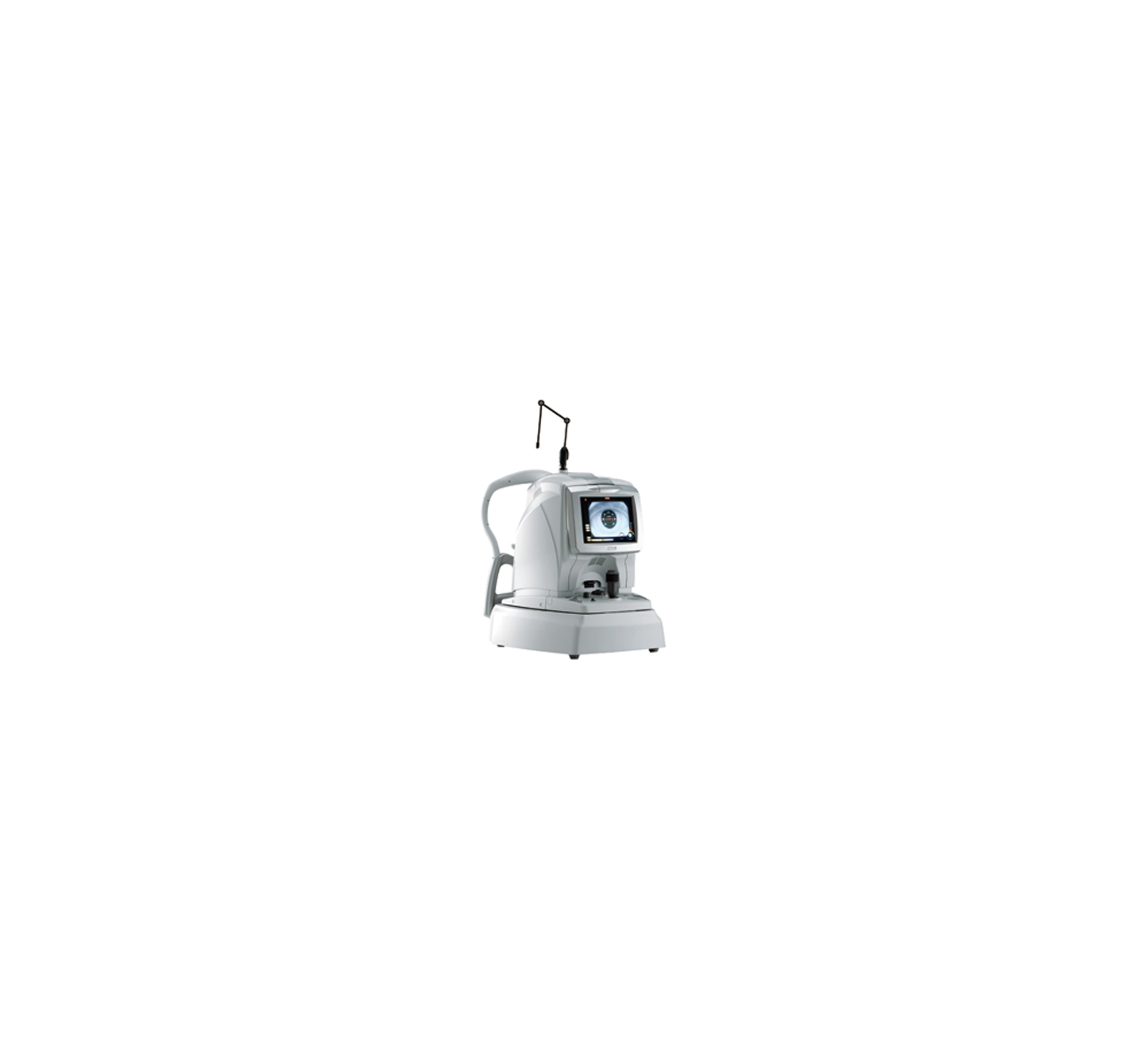
- High speed scan
- High resolution image of OCT & SLO
- Imaging Modes: Scanning Laser Imaging
- Imaging Capabilities: Fundus Camera, Wide-Field Imaging, Retina Thickness, Retinal Mapping, RPE Deformation, Corneal Thickness Mapping (optional), Corneal Curvature Mapping (optional)
- Glaucoma Measurements: TSNIT Analysis, Normative Data Comparison, Optic Disc, RNFL Mapping, RNFL Analysis, Ganglion Cell Complex
The RS-3000 Advanced from Nidek delivers high definition allowing the easy detection of subtle fundus structures and pathology. The model includes repeatable follow up scans of reassuring accuracy using Nidek's tracking and vessel recognition technology, and normative database to aid in diagnosis and comparison of images.
Yes
Not specified
Fundus Camera, Wide-Field Imaging, Retina Thickness, Retinal Mapping, RPE Deformation, Corneal Thickness Mapping (Optional), Corneal Curvature Mapping (Optional)
Not specified
TSNIT Analysis, Normative Data Comparison, Optic Disc, RNFL Mapping, RNFL Analysis, Ganglion Cell Complex
Scanning Laser Imaging
Real-Time Video, 3D Reconstruction
Powercord
—
Not specified
—
- Non-invasive imaging with Retro Mode and FAF
- Field of view is 40° (24x32) 60°, (36x48) -- with non contact wide field lens
- Focus range is -15 to +15 diopters spherical, increments of .5dpt
- Optical resolution is 16 to 20 µm
- EMR compatible
- Pupil diameter is 2.5mm or larger
- Frame rate is 26Hz
- Image Modes: Fluorescein Angiography (FA), ICG Angiography (IA), Fluorescein Angiography (FA) + ICG Angiography (IA), IR reflectance (IR), Blue Reflectance, Green Reflectance, Red Reflectance, Retro Mode, Ring Aperture, Differential Contrast Ophthalmoscopy (DCO)
- Laser Sources: ICG Excitation, Infrared (IR) Reflectance: Laser 790nm, FAG Excitation, Blue Reflectance: Laser 490nm, Green Reflectance: Laser 532nm, Red Reflectance: Laser 660nm
- Light sources: Red Laser, internal 2x2 LED
The F-10 confocal digital ophthalmoscope captures every detail of the retina and choroid, providing high-contrast images with outstanding quality.
Yes
Yes
—
Yes
—
Powercord
—
Yes
—
—
—
—
—

- Providing a comprehensive solution for retina and glaucoma analysis
- Accurate image capture with a SLO-based eye tracer
- Selectable OCT sensitivity that allows acquisition of B-scan images through media opacities
- Tracing HD for accurate averaging of up to 120 image
- Glaucoma analysis with wide-area normative database 9 x 9 mm
- High resolution AngioScan OCT-Angiography image
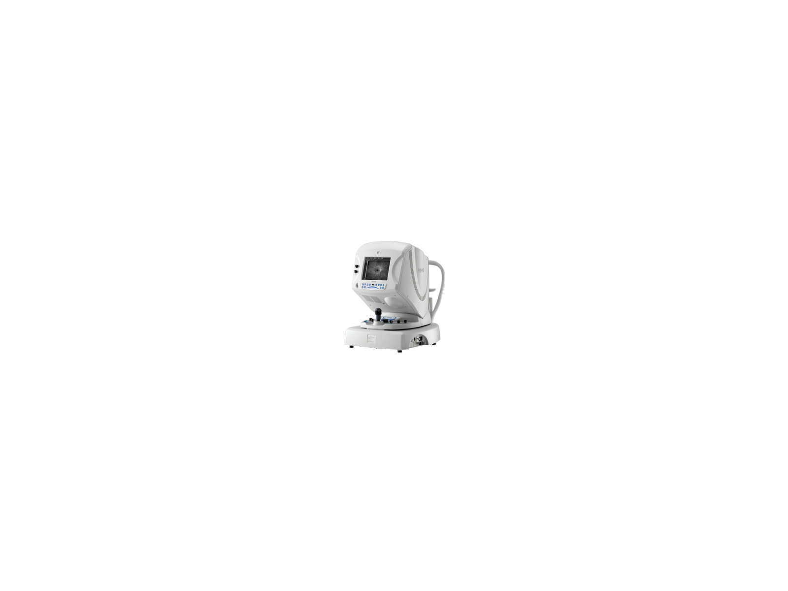
The RS-3000 Advance 2 incorporates a scanning laser ophthalmoscope and is designed for comprehensive imaging and analysis of the retina and glaucoma. Enhancements from the previous model include 85,000 A-scans/s, 12 x 12 mm auto panorama imaging of OCT angiography, and increased image quality.
No
Yes
Fundus Camera, Scanning Laser Imaging
Yes
OCT Angiography+Microperimetry, Normative DB+Microperimetry, Thickness Map DB+Microperimetry, Retinal mode, Choroidal mode
—
Not specified
—
Powercord
—
Not specified
—
—
- Auto-alignment, auto-focus, auto-exposure, and auto-capture
- Imaging modalities include color, IR, and red-free
- Infrared, live viewing of a wide field (out to 110°) is possible using the programmable internal fixation target.
EIDON combines the advantages of SLO with the fidelity of true color imaging. It provides superb image quality, 60° field in a single exposure, a unique, live, confocal view of the retina, three different imaging modalities and dilation-free operation, in a versatile system. The device is operated via a tablet with a multi-touch, high resolution, color display; it works with a dedicated software application and operates as a standalone unit. A joystick is provided when manual operation of the device is desired.
Yes
Yes
—
Yes
—
Color, IR, and red-free
—
Powercord
—
Standalone
—
Not specified
—
—
—
Privacy | Terms & Conditions Ⓒ 2026 Beye, LLC. All rights reserved.
