- Product Listings
- Compare Products
- Calculators
- Product Tours
- Expert Reviews
- Our Experts
- Company Directory
- About Beye
- Contact Us
Ⓒ 2026 Beye.com. All rights reserved.
This content is intended for health care professionals and providers only. The information contained on Beye.com, including text, graphics, images, and interactive activities, is for informational purposes only, and is not intended to be a substitute for professional medical advice. Beye LLC, via its Editors and Publisher, accepts no responsibility for any injury or damage to persons or property occasioned through the implementation of any ideas or use of any product described herein. Although great care is taken to ensure that all information is accurate, it is recommended that readers seek independent verification of advice on drugs and other product usage, surgical techniques and clinical processes prior to their use. References made in article may indicate usage of medical equipment or drugs at dosages, for periods of time, and in combination not included in the current prescribing information. Inclusion of advertising materials on the website thereof, does not constitute and representation or guarantee by Beye LLC of the quality of such products, or of the claims made.
Compare Surgical Microscopes
22 Products
reset all
At-a-Glance
Description
FDA
CE Mark
Type
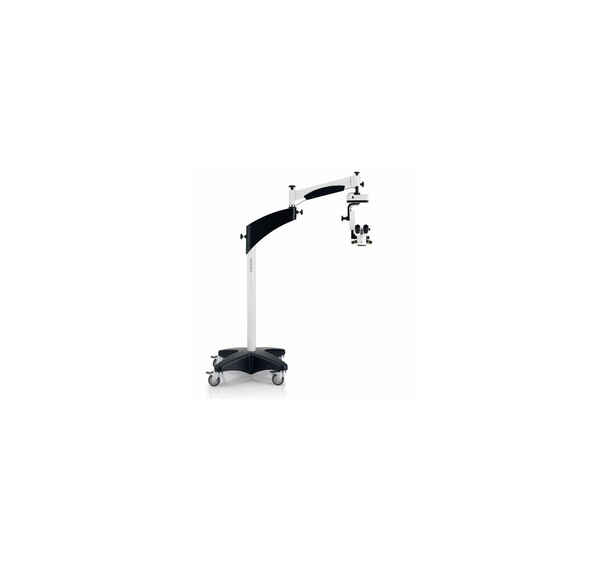
- LED-illumination without fiber optics cables for direct and instant Red Reflex
- Upgradeable XY-unit
- Compact, long-reach stand system allowing high degree of positioning freedom
- LED-illumination; Homogenous, coaxial, integrated red reflex illumination
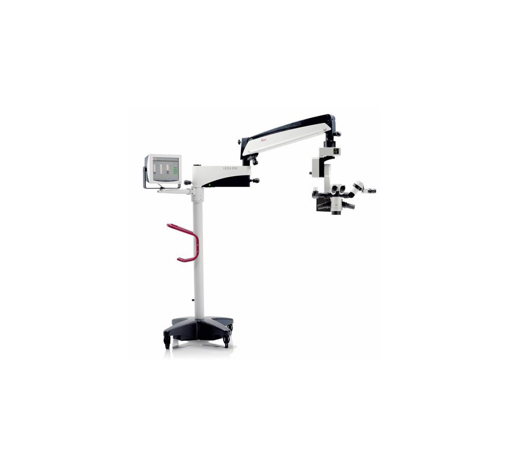
The Leica M220 F12 microscope is used for routine ophthalmic surgery, with 5-step APO-chromatic magnification changer and focus, contrast-rich image, and LED-illumination. The microscope is compact in size and long in reach for positioning flexibility.
—
—
—

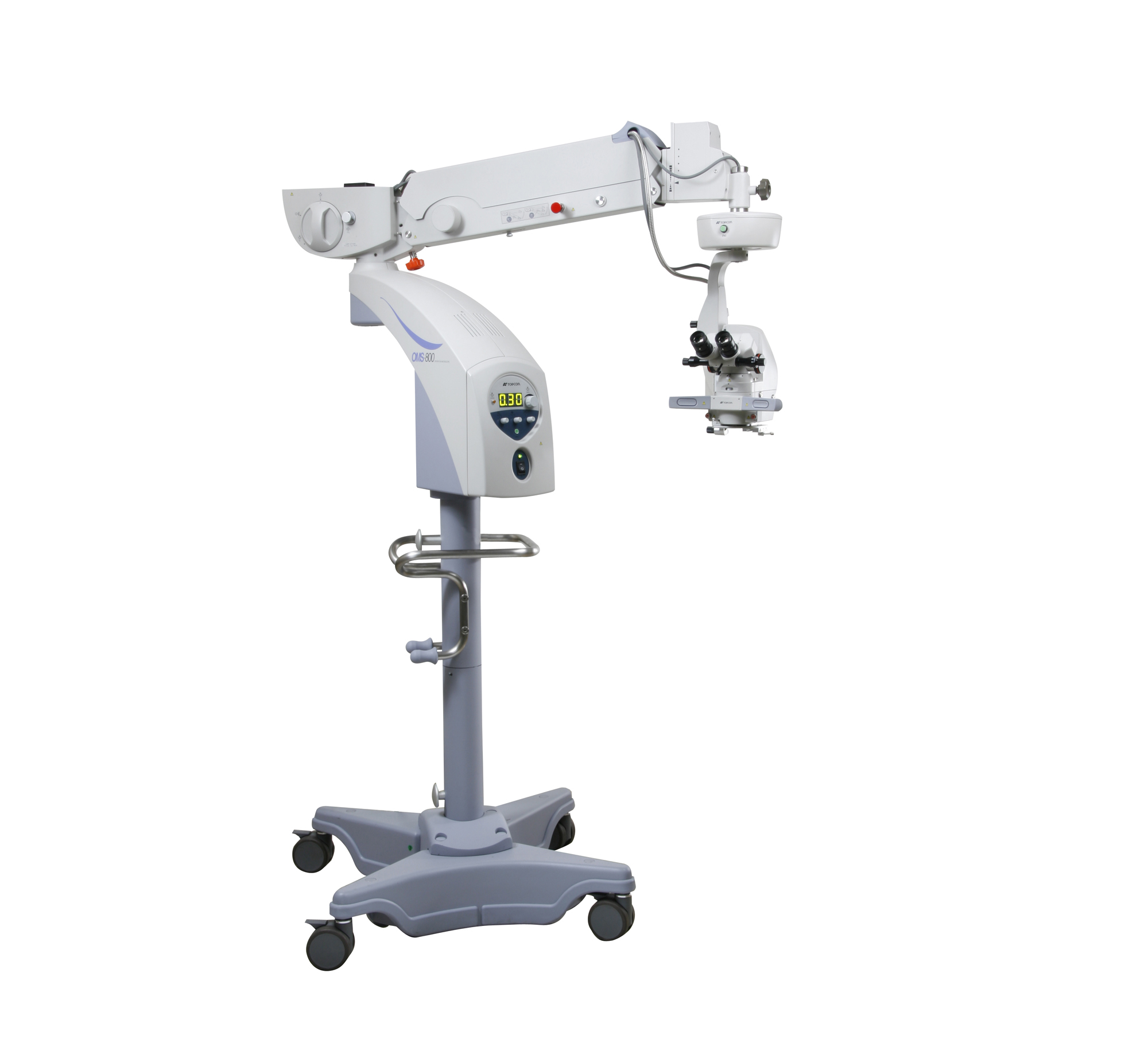
- Electromagnetic locking function enables the surgeon to quickly move the optical head to the preferred position
- The dual-motorized focusing mechanism is a fast focusing system that allows the optical to be quickly elevated during surgery
- Variable angle binocular tube
- XY translator
- A yellow filter and 3-directional illumination ensure a versatile illumination system
- Binocular Tube: Parallel binocular tube, variable angle
- Eyepiece: 12.5x
- Magnification Changes: Continuous zoom
- Total Magnification: 4.2 - 21x
- 100W halogen lamp light source
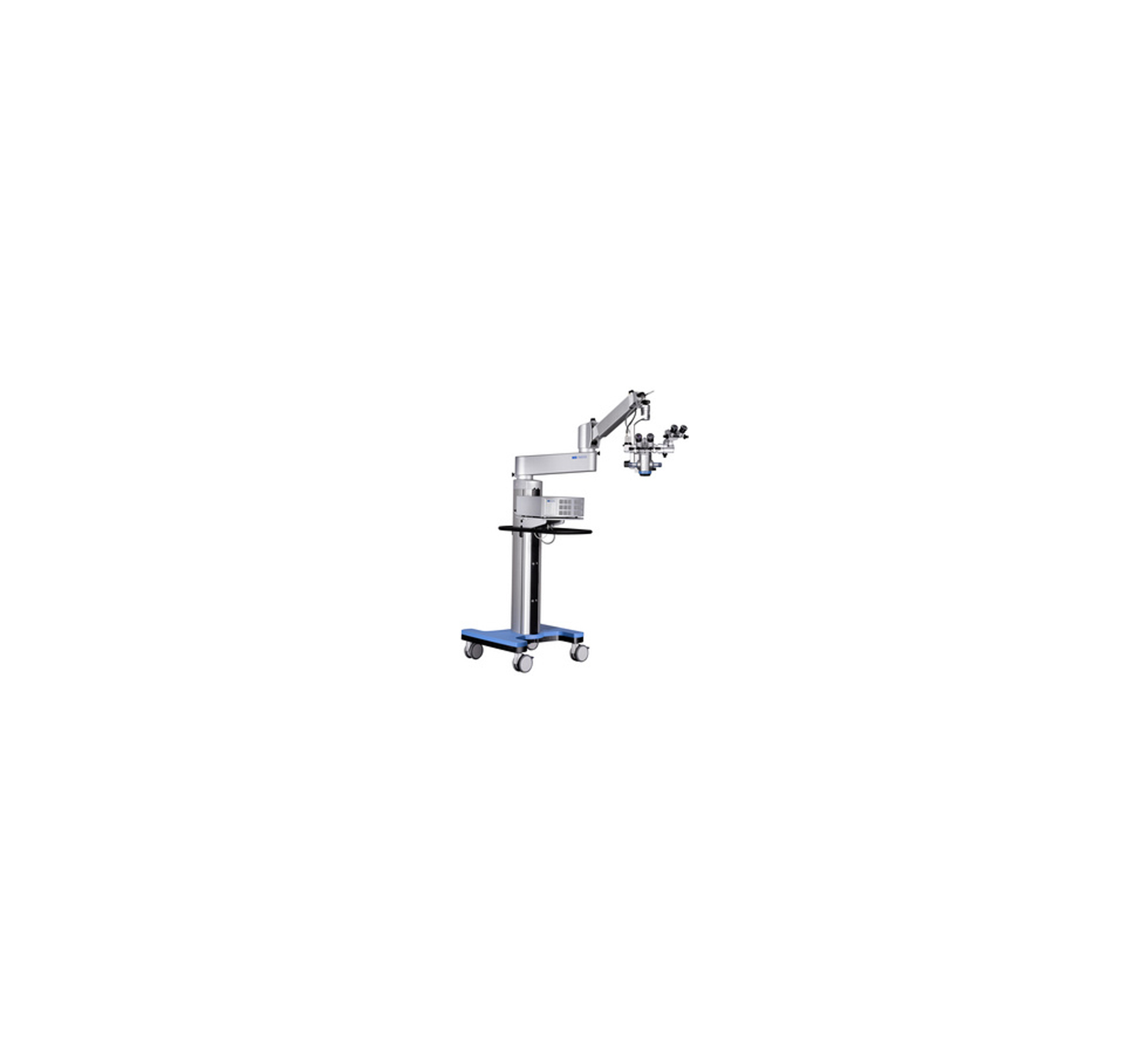
Similar to the standard model, the OMS-800 PRO Operation Microscope can be used for vitreoretinal and cataract surgery with a variable angle binocular tube and XY translator incorporated. In addition to the standard features, this microscope incorporates electromagnetic brakes and a dual-motorized focusing mechanism.
—
—
—
- Stain proof coated objective lens
- 3-directional illumination system (+/-2 & 4 degrees) to ensure versatility
- Yellow filter for protection against phototoxity
- XY-Translator incorporated
- Multifunctional footswitch
- Binocular Tube: Parallel binocular tube, variable angle
- Eyepiece: 12.5x
- Total Magnification: 4.2 - 21x
- 100W halogen lamp light source
The Topcon OMS-800 Standard can be used for vitreous and cataract surgery. It has a variable angle binocular tube and an X-Y translator incorporated, with flexible set up in the operating room and 3-D, versatile illumination.
—
—
—
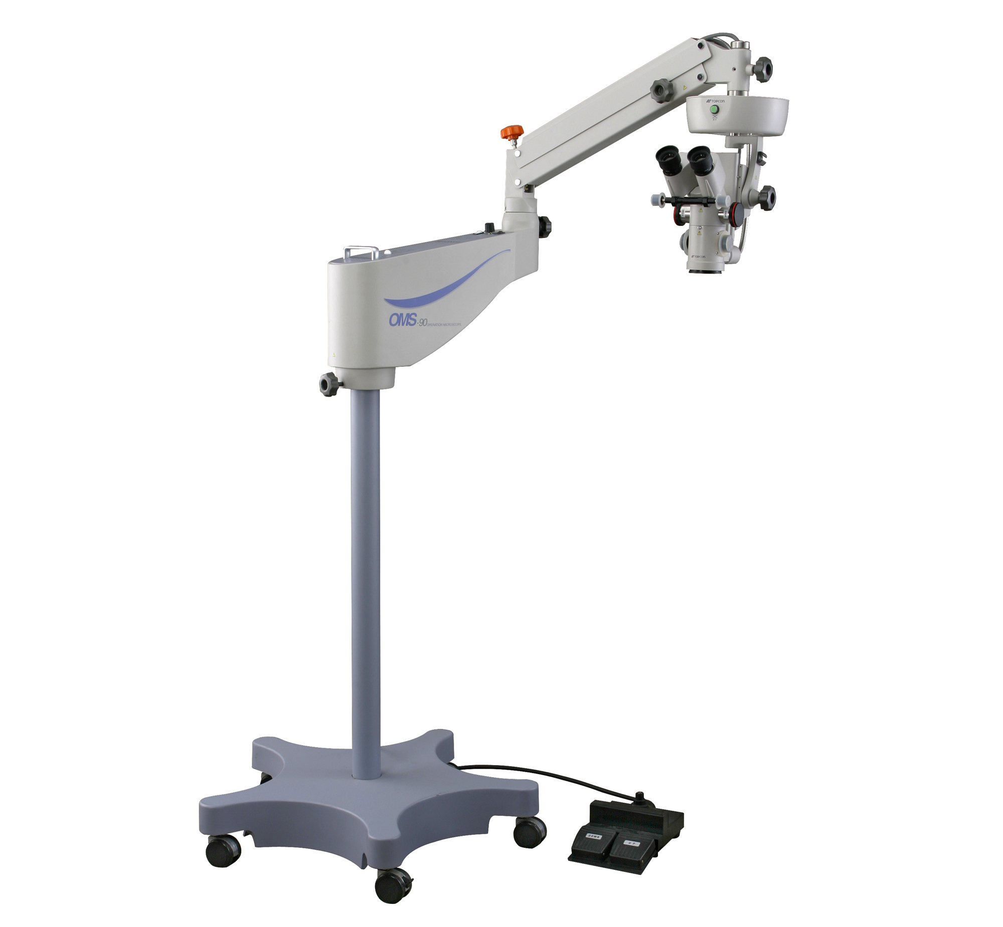
- Stain-proof coated objective lens
- Compact base: 29.92 in. x 29.92 in. (760 mm x 760 mm)
- Binocular Tube: Parallel 45° inclined binocular tube
- Eyepiece: 12.5x
- Magnification Changes: 5-step magnification changer (drum type)
- Total Magnification: 3.4x, 5.3x, 8.5x, 13.6x, 21.2x
- Field Of View: 66.3 mm, 42.3 mm, 26/3 mm, 17.1 mm, 10.3 mm
- 12V/100W halogen lamp light source
The OMS-90 Operation Microscope by Topcon Medical Systems is used for versatile microsurgery with coaxial illumination, a PD adjustment knob, and five-step magnification.
—
—
—
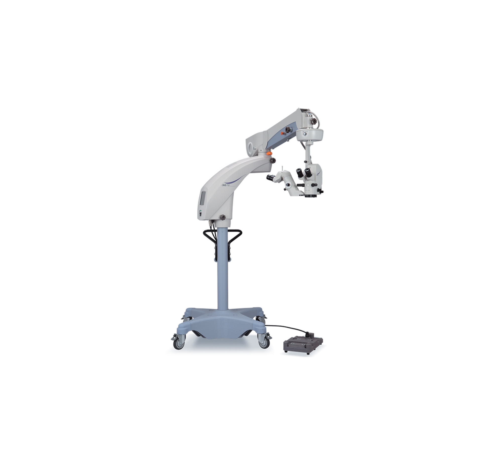
- Optical performance aided by apochromatic optical system for bright, vivid images and natural color with quality red reflex
- Reduced low illumination levels and UV/IR cut filters provide protection from phototoxicity
- Comfortable observation angle with fixed and variable inclination adjustable binoculars offered
- Compact base and longer arms allow easy mobility and storage in limited space
- XY translator and touch panel LCD monitor
- Binocular Tube: Fixed Inclined 45° or Variable Inclined Angle adjustable from 45° to 90° apochromatic optics
- Eyepiece: 12.5x
- Magnification Changes: Continuous zoom
- Total Magnification: 4.2 - 21x
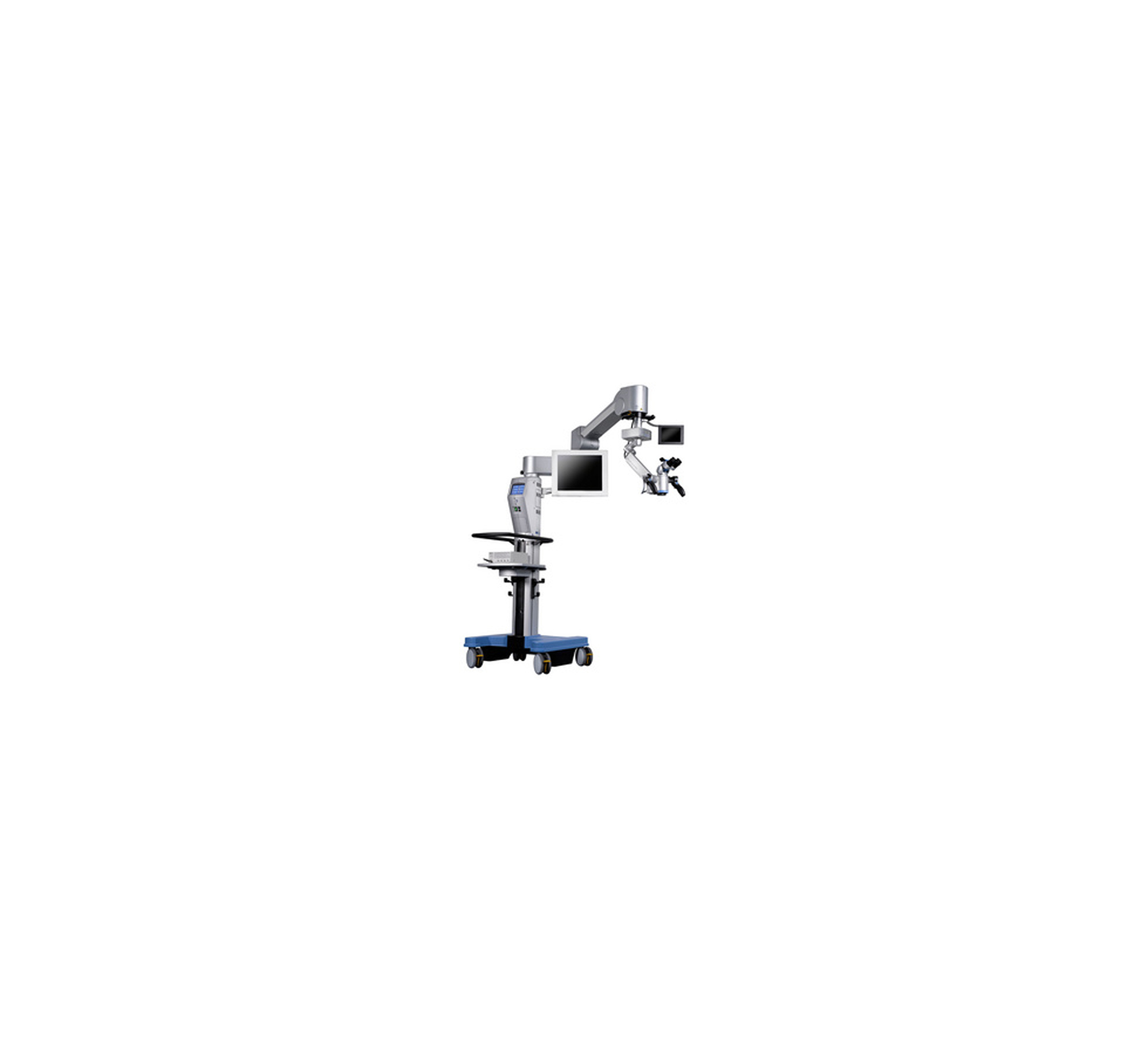
The OMS-710 Operation Microscope is a standard microscope with coverage of anterior and posterior segments. It is said to be simple to operate and contains low illumination levels for protection.
—
—
—
- Provides high stereopsis and bright halogen illumination
- Imaging from anterior to intraocular structures are produced and viewed with distinct precision
- Adjustment of height is made by counter-balance and the fine adjustment of microscope is motorized by footswitch operation without using hands
- Binocular Tube: Inclined and straight
- Eyepiece: 10x
- Magnification Changes: Manual 5-steps changer (drum type)
- Total Magnification: 2.6x, 4.1x, 6.4x, 10.0x, 15.8x
- Field Of View: 69 mm, 44 mm, 28 mm, 18 mm, 11.4 mm
- Focusing Speed: 36 mm by motor (fine adjustment)
- Working Distance: 158 mm
- Interpupillary Distance Adjustment: 52 - 78 mm
- Halogen illumination, 15V/150W halogen lamp
- Field Of Illumination: 48 mm
- Catathermic and green filters
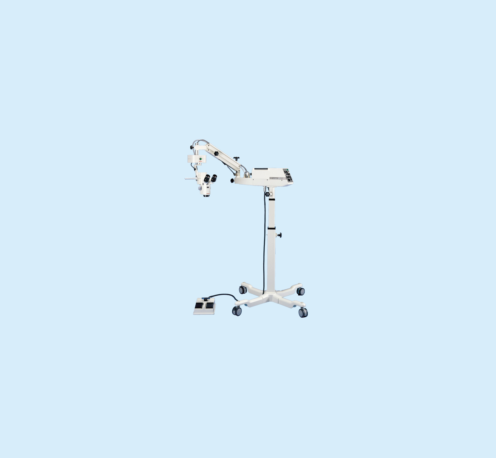
Designed for microsurgery, especially for the eye, the Shin-Nippon OP-2 Operation Microscope includes a five-steps magnification changer, footswitch operation, and sterilization protectors on the change knobs and positioning handle.
Yes
Yes
—
- Can be used on instrument stand to eliminate dedicated room
- Three-step magnification drum and motorized focusing
- Offers a variety of optional accessories
- Documentation is also possible with available video and 35mm camera kits
- Binocular Tube: 45° inclined with converging optics
- Eyepiece: 12.5x
- Magnification Changes: Built-in manual 3-step changer
- Total Magnification: 4.6x, 7.7x, 12.3x
- Field Of View: 43.5 mm, 26 mm, 16.3 mm
- Coaxial adjustment by optical light-guide system
- 15V/150W halogen lamp light source
- Filters include cobalt blue, red-free, heat-absorbing, UV retina shield
- Base size: 23.6 in. x 22.0 in. (600 mm x 560mm), Height: 59.84 in. (1520 mm)
- Weight: 132 lbs. (60 kg)
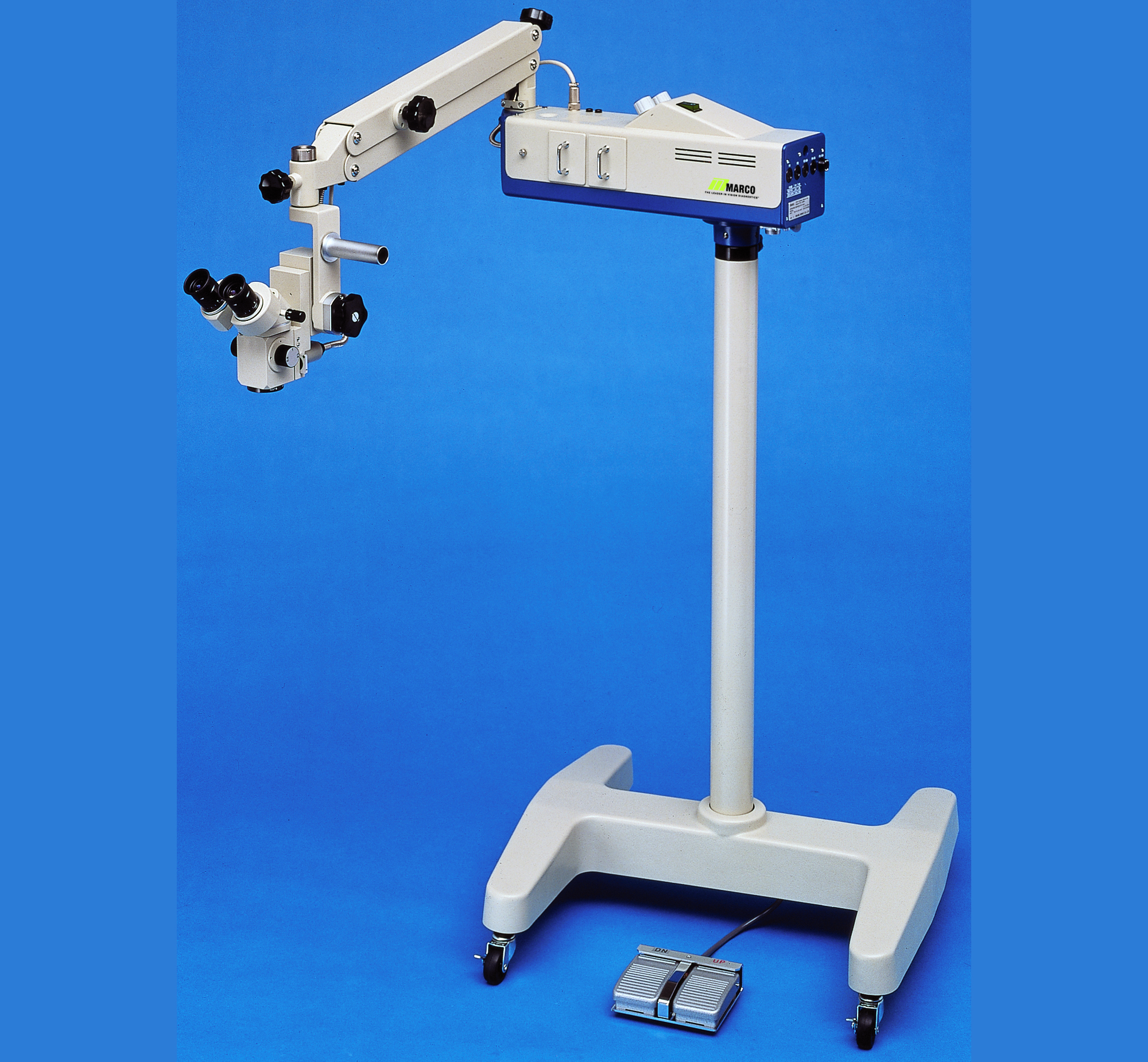
The Marco Surgiscope 3 is a microscope dedicated for in-office surgical procedures, utilizing a simple and compact design. The microscope is equipped with motorized focusing and uses a dual halogen lamp illumination system.
—
—
—
- Leica APO OptiChrome M844 Optics provide high resolution to see small anatomical details
- QuadZoom technology gives surgeon and assistant 100% of the illumination and same magnification
- Exclusive direct illumination system offers clarity, contrast, and color at safe low-light levels for fatigue-free viewing and patient safety
- Two-in-One display control unit
- Wide range of ergonomics to ensure comfort with independent choice of wide-angle observation accessories
- Binocular Tube: APO-chromatic corrected optics
- Eyepiece: 10x
- Magnification Changes: APO-Zoom 6:1, motorized, with 4 separate beam paths
- Total Magnification: 3.5 - 21x
- Field Of View: 7 - 80 mm
- Two halogen lamp light sources
- F40 is slightly larger and has 4 electromagnetic brakes where as the F20 has 3 friction brakes
The Leica M844 F40 / F20 combines optical quality with new technologies of today's surgical microscopes. The microscope uses APO OptiChrome optics and direct halogen illumination for safe low-light levels, with additional QuadZoom and Double Wing Assistant Bridge technologies.
—
—
—
- Precision Leica optics paired with a dual LED and halogen illumination system for stable red reflex
- Ergonomic design and easy to use, with comfortable vision and posture for the surgeon, and intuitive control for efficient workflow and precise microsurgery
- Total image management through high-definition video for display, documentation, and communication of procedures and cases
- F40 is slightly larger and has 4 electromagnetic brakes whereas the F20 has 3 friction breaks
- Binocular Tube: APO-chromatic corrected optics
- Eyepiece: 10x
- Magnification Changes: APO-Zoom 6:1, motorized, with 2 separate beam paths
- Total Magnification: 3.5 - 21x
- Field Of View: 7 - 80 mm
- Focusing Speed: Focus range: 54 mm, motorized, automatic reset
- Working Distance: 175 mm, 200 mm, 225 mm
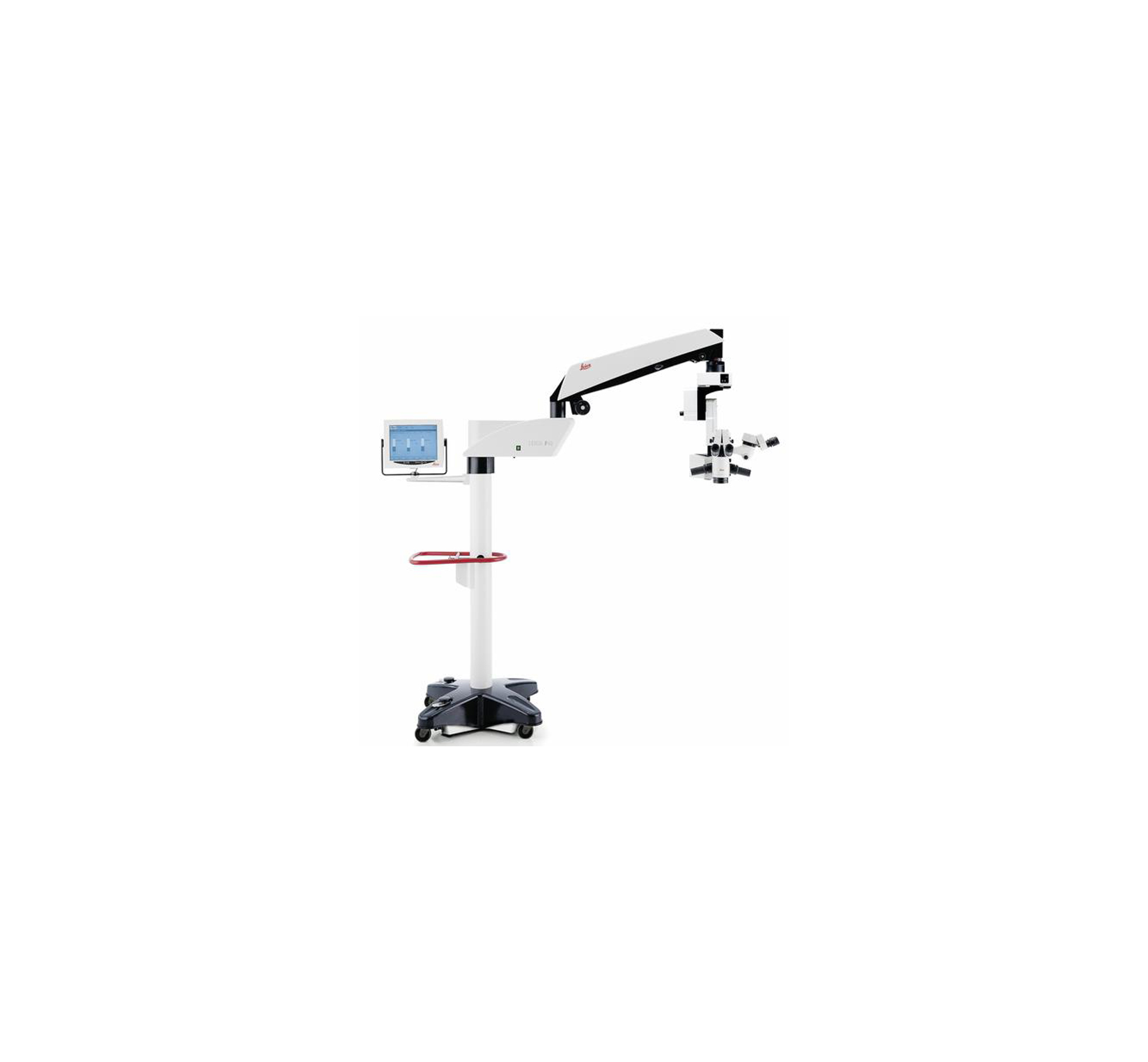
The Leica M822 F40 / F20 surgical microscope, with enhanced red reflex, uses an LED and halogen illumination combination for surgeons performing cataract surgery. High-def video via Leica HD C100 high definition medical grade camera is also included, and the F40 / F20 options serve as interchangeable floor stands for upgradability.
—
—
—
- Leica 800-series APO OptiChrome provides sharp image, natural color, depth of focus, and high contrast for all types of ophthalmic microsurgery
- Leica M820 optics provides added patient safety by allowing the surgeon to use lower levels of illumination
- Double beam stereo illumination creates true, three-dimensional illumination for quick and accurate surgical procedure completion and a stable red reflex
- Binocular Tube: APO-chromatic correct optics
- Eyepiece: 8.33x, 10x, 12.5x
- Magnification Changes: APO-Zoom 6:1, motorized, with 2 separate beam paths
- Total Magnification: 3.5 - 21x
- Field Of View: 7 - 80 mm
- Working Distance: 175 mm, 200 mm, 225 mm
- Direct Halogen Illumination system; 800-series double beam illumination system; stable red reflex
- Field Of Illumination: 4 - 35 mm
- IR-barrier and UV-barrier filters
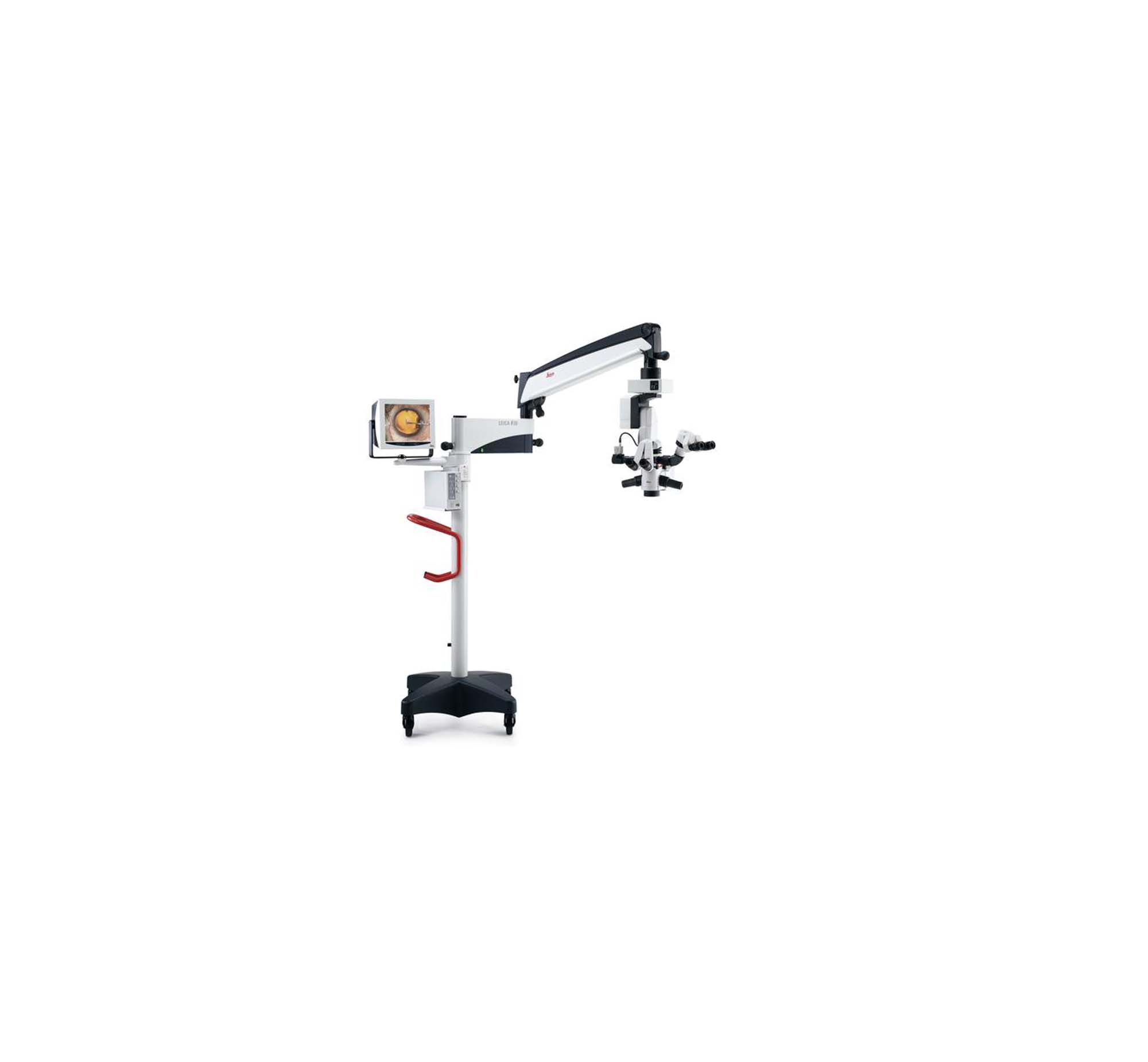
Designed for the ophthalmic market, the Leica M820 F40 / F20 features 800-series APO OptiChrome optics and Direct Halogen Illumination system for natural color, depth-of-field, and a stable red reflex to provide detail recognition. The floor stand has a small base, long reach, and mechanical brakes for easy setups in limited space.
—
—
—
- Leica's direct halogen illumination system allows the surgeon homogeneous illumination to see images at low light levels, and provides a stable Red Reflex
- Compact, light and easy to move with precision bearings and long swing-arm
- Binocular Tube: +/-5° fine adjustment inclining mechanism
- Eyepiece: 8.33x, 10x, 12.5x
- Magnification Changes: 6:1 zoom, motorized, adjustable speed
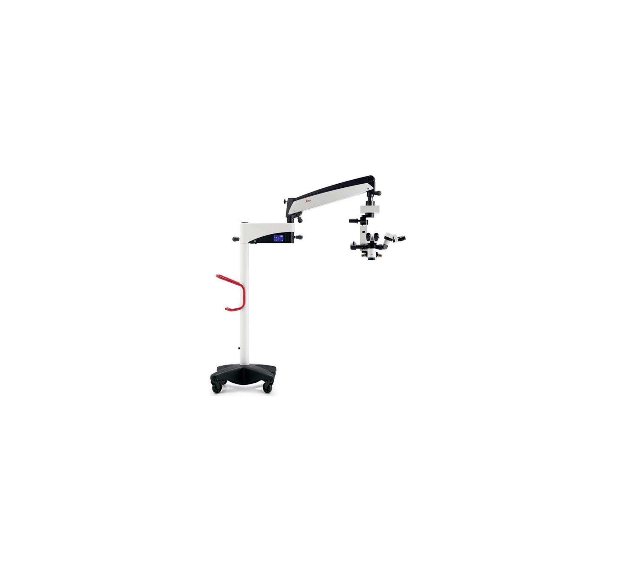
The Leica M620 F20 is an ophthalmic surgical microscope with sharp OptiChrome optics for natural color, depth-of-focus, and high contrast in detail recognition. The microscope includes direct halogen illumination with stable red reflex.
—
—
—
- Can select between Xenon Superlux eye, LED, and established halogen light sources
- Integrated slit illuminator
- RESIGHT 700 fundus viewing system allows surgeon to recognize details of the retina
- Binocular Tube: Apochromatic optics, invertertube
- Eyepiece: 10x
- Magnification Changes:Electric/motorized, focus range: 70 mm
- Total Magnification: 0.4 - 2.4x
- Blue blocking (all light sources), HaMode (Superlux Eye), HaMode (LED fiber optic), and 25% gray (LED fiber optic) filters
Used for cataract and retina procedures, the OPMI LUMERA 700 and RESCAN 700 has a wide range of customization options. The microscope utilizes the Stereo Coaxial Illumination (SCI) system for red reflex and image quality.
—
—
—
- 3D vision and depth perception features
- High comfort due to zoom optics, electromagnetic brakes, and X-Y coupling
- Choice of halogen or LED illumination
- Binocular Tube: -8° to +100° inclination angles with apochromatic optics
- Eyepiece: 10x (160° inclinable eyepiece head)
- Bluetooth wireless footswitch
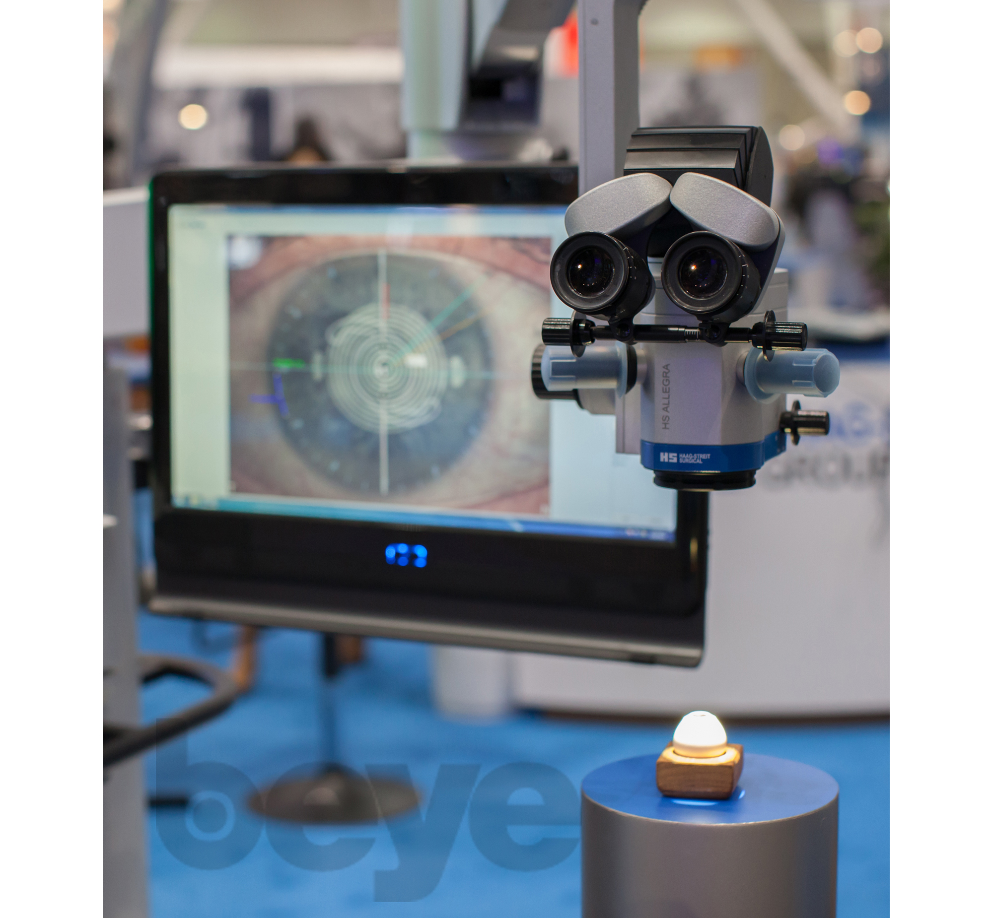
The ALLEGRA 900 is easy to move and stable in the working position, with a newly designed electromagnetic brake recommended for surgical centers. It is very compact, and equipped with a 25 mm stereo base for 3D vision. The microscope also includes red reflex enhancer and motorized zoom and focus.
—
—
—
- Large stereo base to enhance depth perception
- High optical quality
- Fast change of magnification in defined steps
- Flexible movement and positioning with mechanical brakes
- Binocular Tube: -8° to +100° inclination angles with apochromatic optics
- Eyepiece: 12.5x (fix 60°)
- Magnification Changes: 5 step magnification changer
- Total Magnification: 3.3 - 30.3x
- Field Of View: 7 - 64 mm
- 15V/150W halogen lamp, 12V/50W LED light source
- Field Of Illumination: 3 - 60 mm
- Filters include:
- Spot and minispot diaphragms
- Daylight filter
- Blue filter
- Green filter
- Softlight filter
- UV protection filter
The ALLEGRA 90 provides sharp and contrast images at any magnification through tilt and light management, and is recommended for surgical procedures where speed and flexibility are decisive factors, as well as day clinics. A large, 25 mm stereo base provides depth perception.
—
—
—
- 3D vision and depth perception features
- High comfort due to zoom optics and X-Y coupling
- Integrated camera reduces length of the microscope, supports ergonomic positioning, makes it easier to clean
- Designed to rotate cardanically around center of gravity in all directions, requiring only minimal force
- Binocular Tube: Apochromatic optics with fixed inclination angles
- Eyepiece: 10x (160° inclinable eyepiece head)
- Total Magnification: 4.3 - 25.7 x
- Field Of View: 8.2 - 49 mm
- Halogen or LED light source
The ALLEGRA 590 is said to be specialized for dual use in ophthalmology and ENT with depth perception and 3D vision. The microscope is permanently balanced, with flexible rotation, mechanical breaks, and an integrated video camera. The apochromatic optics provides a sharp and contrast image at any zoom setting.
—
—
—
- Intraoperative live OCT
- Individual configuration exactly for your needs and ergonomic demands
- High optical image quality
- Movability and precise positioning
- Large stereo base to enhance depth perception
- Binocular Tube: Coarse -70° to +90°, fine +/-10° inclination angles with apochromatic optics
- Eyepiece: 10x (Inclinable eye piece head 200° with wide angle oculars)
- Total Magnification: 3.9 - 23.2x
- Field Of View: 9.0 - 54.2 mm
- Halogen or LED by clear zoom illumination
- Wireless bluetooth footswitch
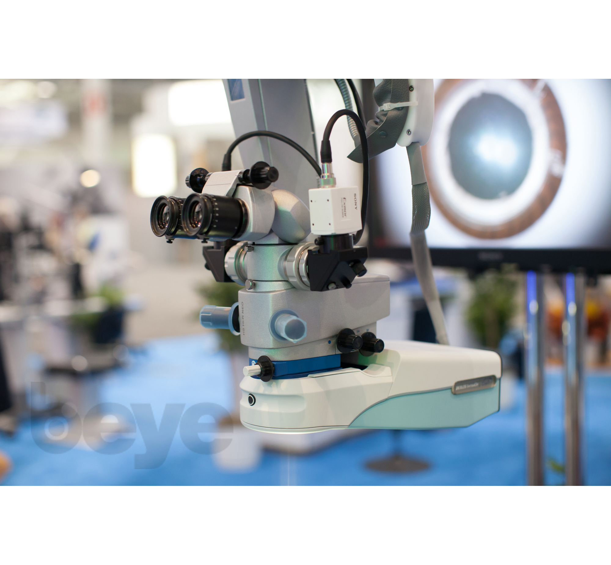
The Hi-R NEO 900 microscope provides high optical visualization for surgical procedures requiring vertical observation, allowing for depth perception through a large stereo base and clear zoom illumination.
—
—
—
- Fatigue-free surgery with 10x21mm visual field
- Optimized halogen-illuminated system
- Optimized red reflex
- User-friendly lamp replacement with two onboard lamps allows the lamp to be changed instantly without interruption
- Built-in, rotatable 7" color TFT LCD control panel
- Binocular Tube: 5 degree to 35 degree tilt
- Eyepiece: For glasses wearers 10x/21mm
- Magnification Changes: Zoom 6:1, motor-driven: variable speed
- Total Magnification: 3.1x (min) to 18.7x (max)
- Field Of View: 68.0mm (max) to 11.1mm (min)
- Field Of Illumination :70mm
- Filters: IR protective filter fixed, 420 UV protective filter fixed, 475 UV protective filter (optional)
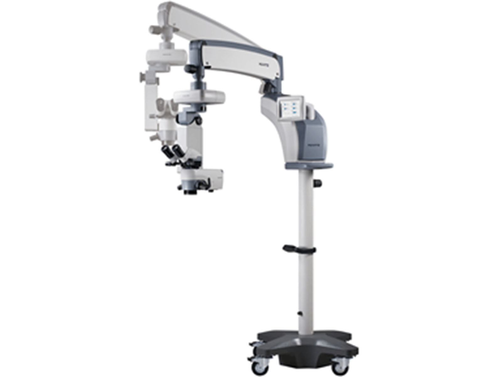
The Huvitz HOM 700 Surgical Microscope features a high resolution optical system, which provides enhanced images even in low-intensity illumination situations enabling high-resolution 3D observations. The device streamlines surgical workflow and maximizes surgery efficiency.
Not specified
Not specified
—
- Touch screen controls both the microscope head and the video camera for central control, intuitive operation, and easy access
- It is small and easy to move, and good for smaller operating rooms
- Ergonomically designed foot control panel allows surgeon to control surgical microscope intuitively and reliably
- Eyepiece: 10x
- Magnification Changes: Motorized zoom system, 1:6 zoom ratio
- Total Magnification: 0.4 - 2.4 x
- 12V/100W halogen lamp light source
The OPMI LUMERA i Surgical Microscope is equipped with Stereo Coaxial Illumination (SCI) for red reflex used in anterior segment work and providing detail recognition for cataract/refractive surgeries.
—
—
—
- Comfort of the LED illumination with natural colors and contrast as well as lower energy and maintenance costs
- No harmful IR or UV radiation for the patient
- Built for stability and reliability, system servicing and maintenance is kept to a minimum
- Easy to use and simple mobility
- Binocular Tube: Apochromatic optics with 45° inclined tube
- Magnification Changes: 5-step magnification changer, optionally motorized
- Total Magnification: .4x, .6x, 1.0x, 1.6x, 2.5x
The OPMI 1 FR PRO Surgical Microscope utilizes LED illumination and is geared toward routine procedures for high patient throughput. It was originally developed for areas of rapid economic development, such as the emerging regions of India and Southeast Asia, where there exists a high demand for ophthalmic systems.
—
—
—
- Optimum visualization and contrast with the Stereo Coaxial Illumination (SCI) system
- Instant red reflex
- Integrated DeepView depth-of-field management system to choose between maximum depth of field and optimum light transmission at the push of a button
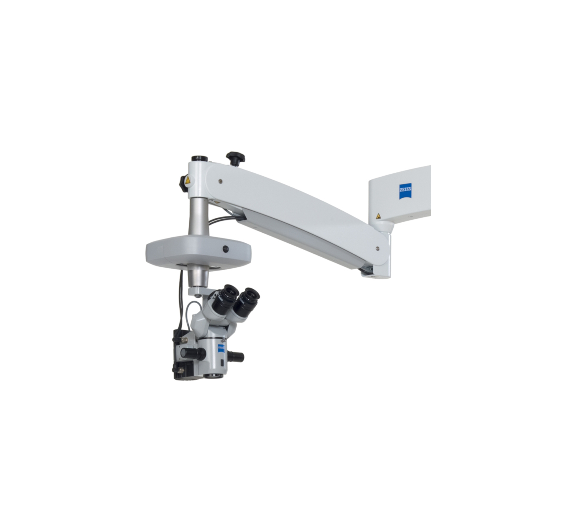
- Versatility in positioning, with choice between ceiling mount suspension or floor stand
- Binocular Tube: Apochromatic optics, 0-180° tiltable tube
- Eyepiece: 10x
- Magnification Changes: Motorized zoom system, 1:6 zoom ratio
- Total Magnification: 0.4 - 2.4 x
- 12V/100 W halogen lamp light source
- UV barrier and Blue blocking filters
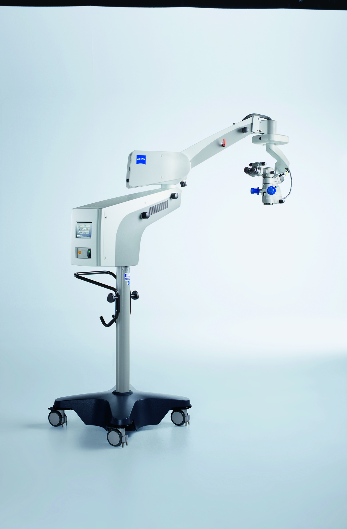
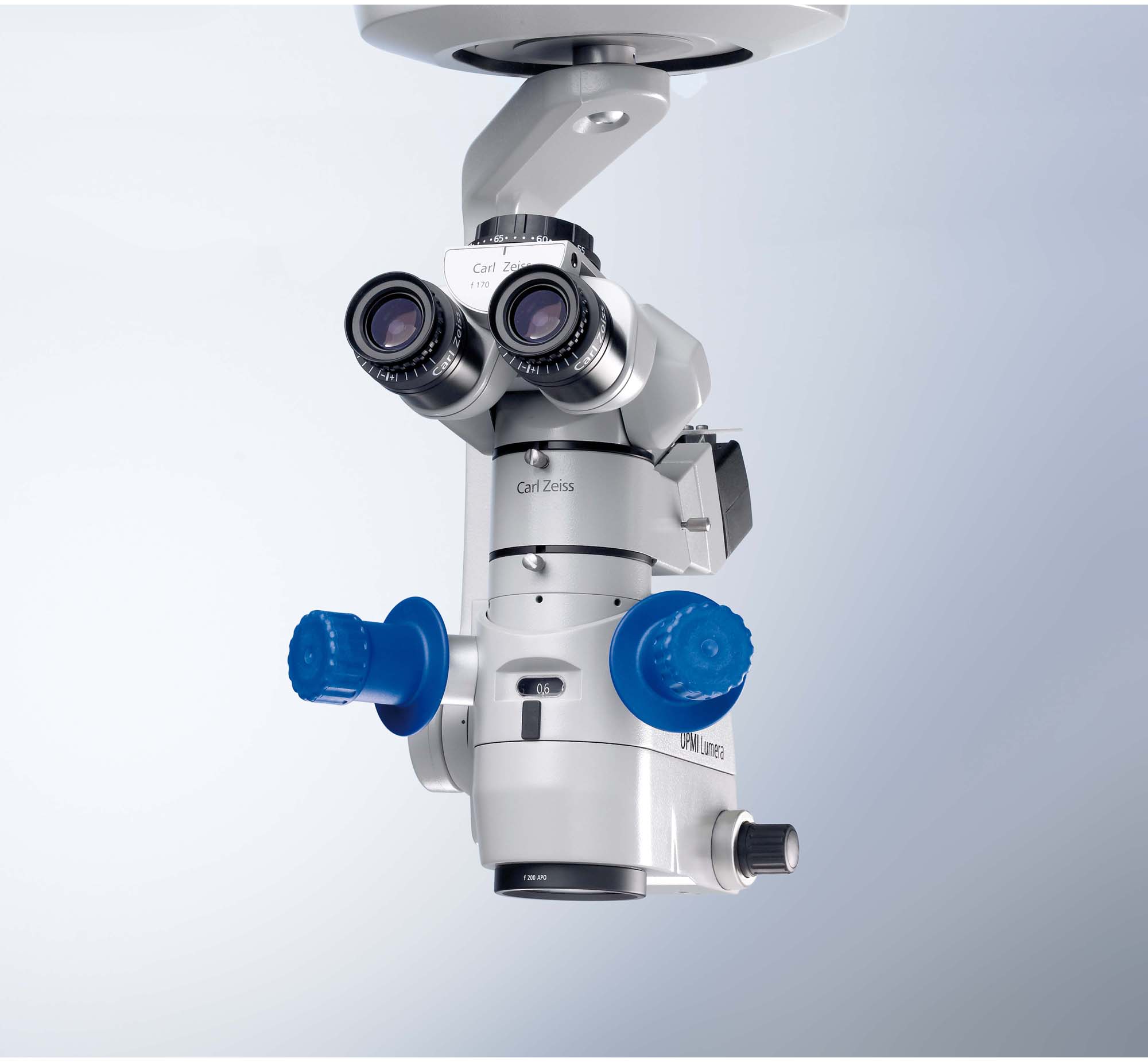
The OPMI LUMERA S7 Surgical Microscope can be suspended using a ceiling mount, or mobile with a floor stand. The microscope utilizes the Stereo Coaxial Illumination (SCI) system for red reflex and image quality.
Yes
Yes
Stereo Coaxial Illumination
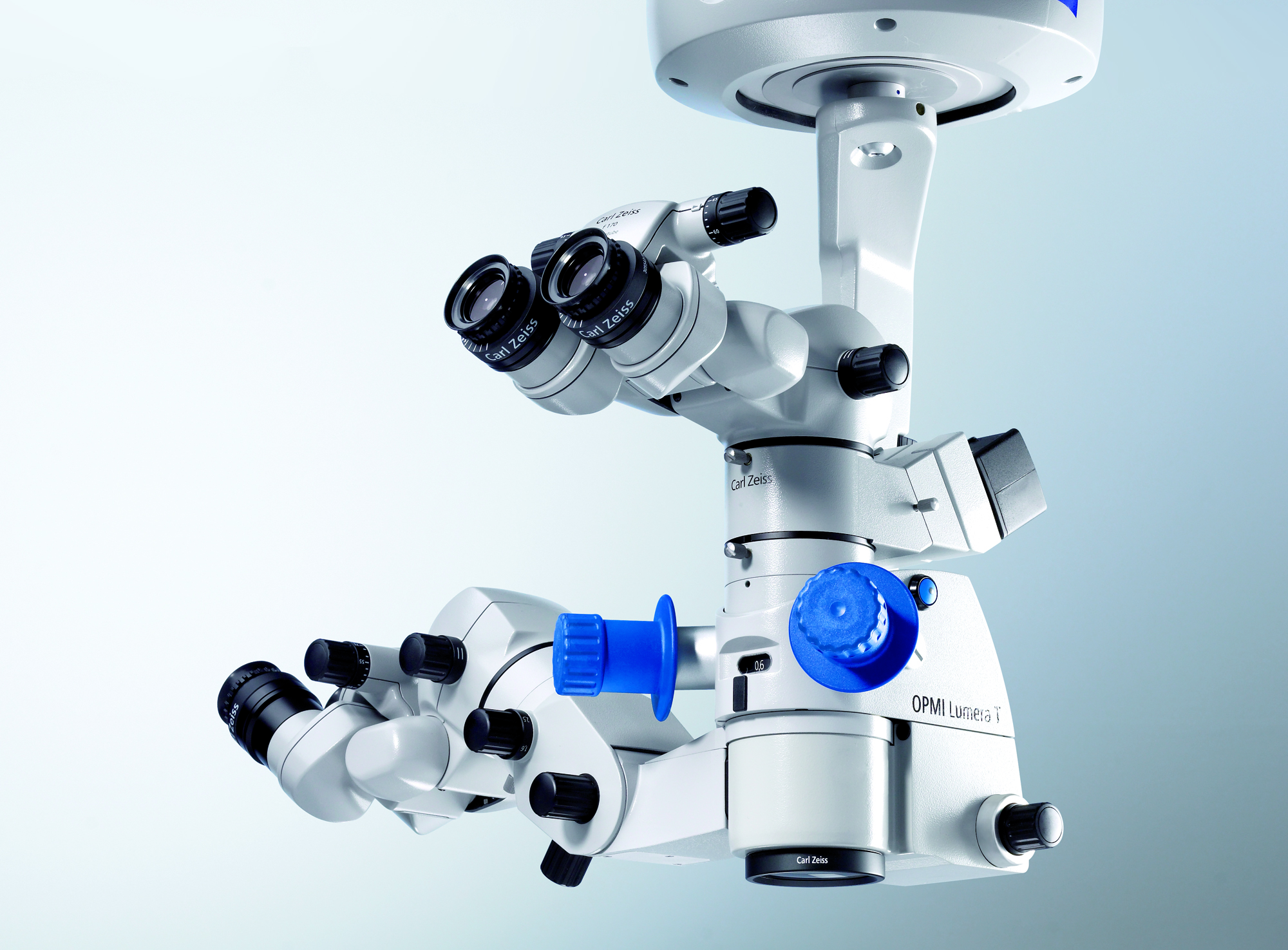
- Stereo Coaxial Illumination (SCI) incorporates illumination technology that optimizes red reflex with heavy detail recognition
- Integrated assistant's microscope with independent focusing and zoom
- DeepView, integrated depth-of-field management system, permits optimization of the microscope image to depth of field or light transmission
- Superlux Eye xenon illumination system offers surgeons white, natural, high contrast image of surgical field
- Binocular Tube: Apochromatic optics, invertertube
- Eyepiece: 10x
- Magnification Changes: Motorized zoom system, 1:6 zoom ratio
- Total Magnification: 0.4 - 2.4 x
- Red reflex illumination and surrounding field illumination, both dimmable
- Superlux Eye Xenon Light Source
- HaMode and blue blocking filters
- Base Size 31.69 in. x 31.69 in. (805 mm x 805 mm); Height 74.02 in. (1880 mm)
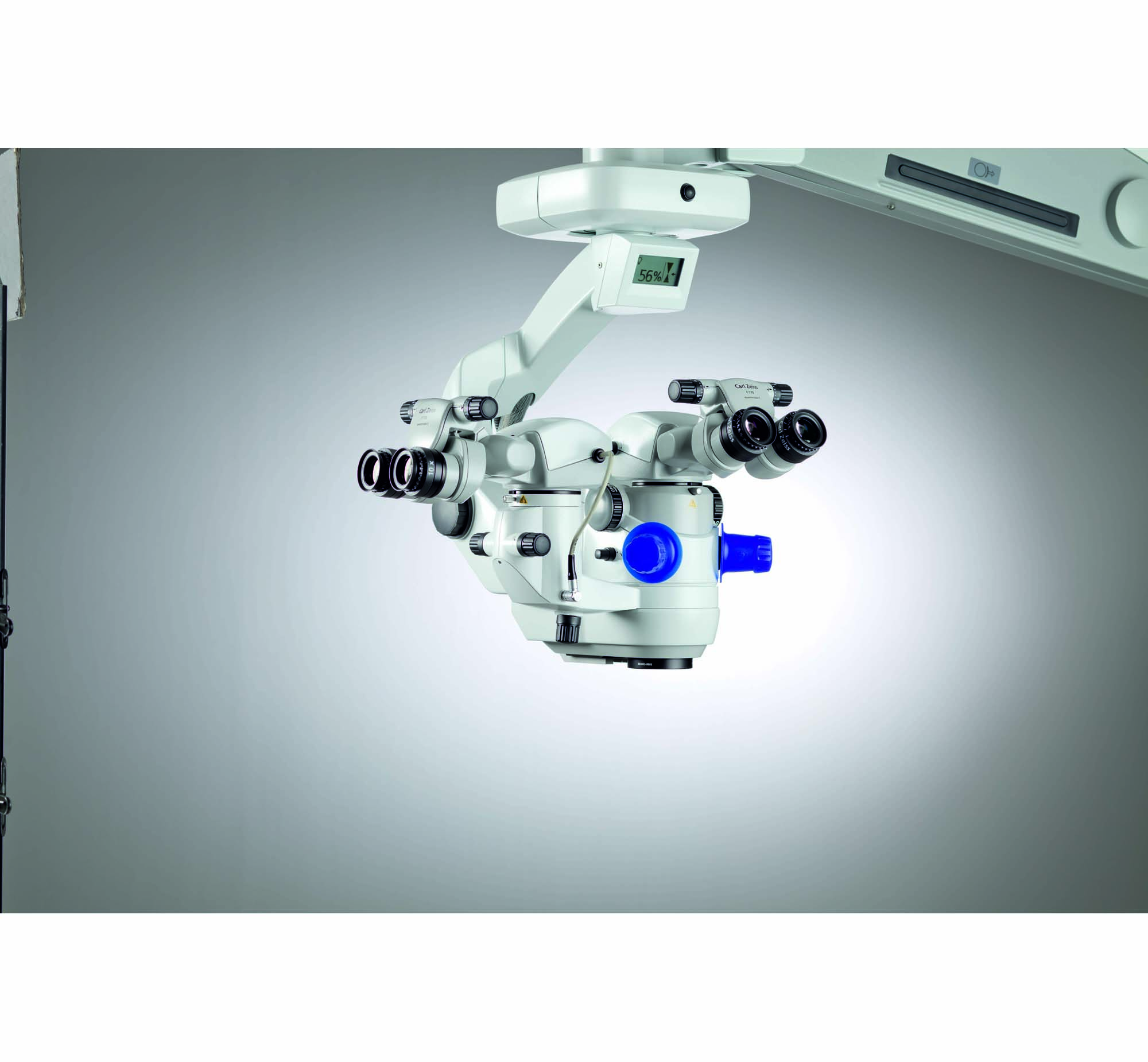
The OPMI LUMERA T Surgical Microscope is equipped with Stereo Coaxial Illumination (SCI) to provide red reflex and detail recognition for cataract/refractive surgeries.
—
—
—
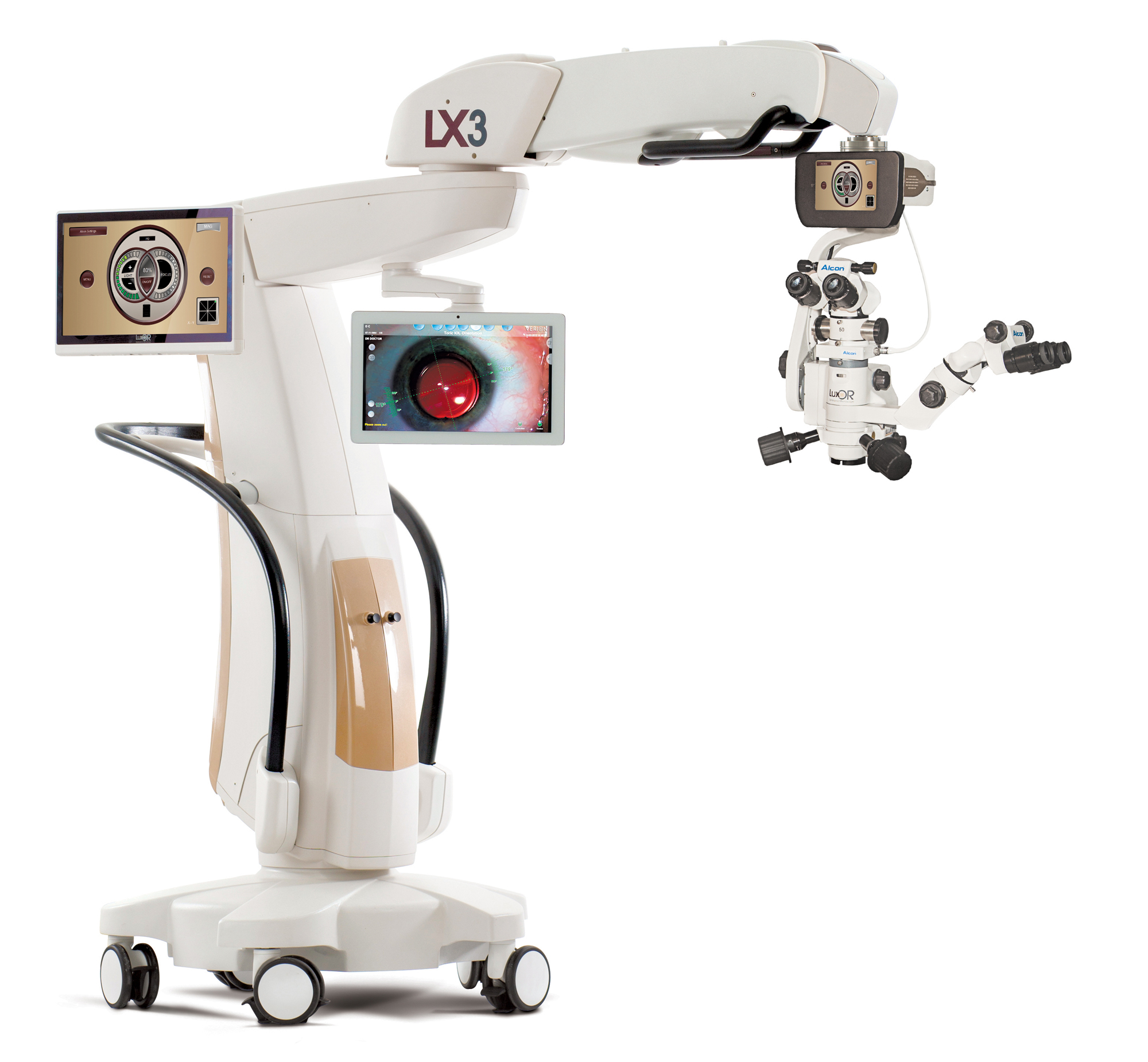
- ILLUMIN-i technology delivers a 6x-larger, highly stable red reflex zone and a greater depth of focus
- LX3 floor stand combines increased mobility and functional design to improve surgeon and staff experience
- Q-VUE Assistant Visualization provides a 3-D stereo assistant scope that takes no light from the surgeon's optical pathway
- Field Of Illumination: 1882.90 mm2 (area of red reflex zone)
The LuxOR LX3 with Q-VUE Ophthalmic Microscope is designed to deliver visualization throughout the cataract refractive surgical procedure, with illumination technology and comprehensive visualization.
—
—
—
Privacy | Terms & Conditions Ⓒ 2026 Beye, LLC. All rights reserved.
