Editorially Independent Content
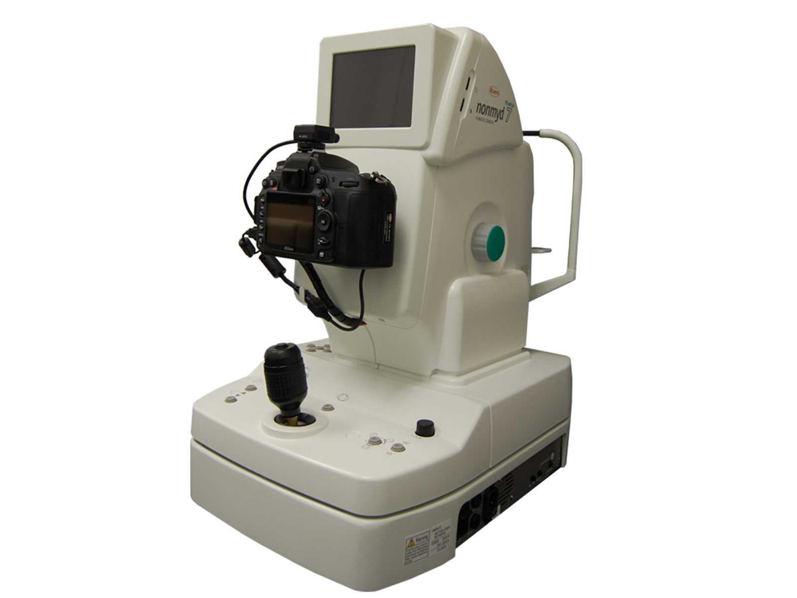
The Kowa nonmyd 7 is a non-mydriatic digital retinal camera that captures high-resolution images at two different optical magnifications. The 45° view offers the standard perspective and the 20° view is a more magnified option for closer examination. An integrated Nikon digital SLR camera captures high levels of clinical detail.
Camera Features
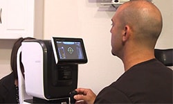
The Kowa nonmyd 7 unit connects to a large secondary monitor and the output is also displayed on the unit during image capture, which simplifies operation. By toggling a few buttons, a user can choose to capture a nasal or temporal bias photo rather than just a central 45° photo. This works well for patients with a temporal lesion or diabetic retinopathy outside the central part of the scan. Additionally, the automated chinrest moves up and down at the push of a button. Technicians have also found the unit very easy to capture high-quality images even on small pupils.
Another useful feature of this particular camera is the ability to be networked to multiple exam rooms. If moving from one exam room to another, staff can access the patient’s retinal scan when they enter the room.
Robust Image Processing and Filter Tools
The VK-2 digital imaging software common to all Kowa retinal cameras is quite robust. One can view chronological scans and easily magnify areas to thoroughly examine the retina or macula. With a few mouse clicks, the measurement tool can identify the size of a lesion or optic nerve. Additionally, one can apply filters to the photos to better diagnose potential pathology. There are red, green, and blue filters as well as a negative for a reverse polarization. The green filter is especially helpful when evaluating a nevus.
Limited but Adequate View
While the 45° and 20° views do not show as much of the periphery as the widefield cameras on the market, the angles do capture most of what is needed in very high resolution.