Editorially Independent Content
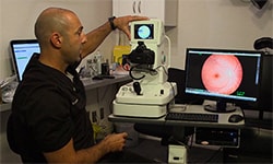
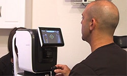
The AcuTarget HD (Visiometrics) is a four-in-one diagnostic and surgical planning instrument that measures depth of focus, light scatter, tear film quality, and identifies ocular landmarks to guide corneal inlay placement. The device was developed by Visiometrics S.L. in collaboration with AcuFocus, Inc.
Four Powerful Measurements The AcuTarget HD measures forward light scatter in the eye, which objectively quantifies quality of vision. This gauge is used to assess if patients have cataracts, and if so, the level of their cataract. It can also be used to evaluate corneal disorders or dysfunction and how they may be affecting quality of vision. The Ocular Scatter Index (OSI) scale ranges from zero to six, where lower scores indicate better quality of vision. A score below 1.2 is considered good. The patient’s measurement is also plotted on a color-coded scale ranging from green to yellow to red. Additionally, the screen shows a simulation of an image on the patient’s retina in comparison to what a perfect optical system would perceive. Both the color-coded scale and retinal image simulation are nice tools to show patients so they can easily understand the quality of their vision.
Another nice feature of the OSI report is the predicted visual acuity measurement. This is different from the visual acuity that you would measure in an exam lane. The value is based solely on the optics of the eye as measured by the instrument. It tells you how high quality the patient’s optical system is, which can help narrow down differential diagnoses to figure out if the vision issue is related to the cornea or the lens, or involves the retina or neurological problems.
The second module assesses tear film quality to understand how a patient’s ocular surface is affecting their quality of vision. During this test, the patient is asked to focus their eye and not blink for 20 seconds. The instrument evaluates how stable the patient’s tear film is during that time. It’s the only diagnostic instrument of its kind that can measure the effects of the tear film on the quality of vision, and ultimately how well a patient sees. A flat line indicates that the tear film is not interfering with vision. The line begins to rise after 5 seconds in patients with a lipid disorder and indicates the degree to which the ocular surface is affecting vision.
The third feature analyzes depth of field, or how well patient can focus from near to far. Again, this module includes visual representations of what the patient sees to help him understand his vision and how it changes over time.
The fourth attribute of the AcuTarget HD, which separates it from the HD Analyzer (Visiometrics), is the software that allows you to analyze the landmarks in the eye – mainly the pupil center and purkinje image. This can help doctors understand where to place the refractive treatments, like LASIK or corneal inlays such as the KAMRA inlay. There is also a post-operative module that measures where the inlay is located relative to these landmarks to inform the surgeon if it is in the optimal position.
Room for Workflow Improvement While there are a lot of advantages to this technology, there are a couple of aspects I would change. First, the instrumentation does not come with a wireless mouse so I highly recommend that you purchase one separately. It makes measuring the patients easier and faster. Secondly, you have to print out each measurement individually. It would be nice to able to print everything out all at once, which is reportedly a feature in the next software generation.
Another disadvantage is that patients must have their ametropia corrected in order for the test to be run correctly. If patients have a pair of glasses, they can wear them during the test. Alternatively, you have to put trial lenses into the instrumentation in order to correct the patient’s vision. Sometimes this can be cumbersome if the patient has a very high prescription beyond -6D or + 6D or an astigmatism.
Overall the AcuTarget HD is a powerful tool that streamlines evaluations so doctors can obtain an objective assessment of how well a patient sees and determine the source of quality issues.