Editorially Independent Content
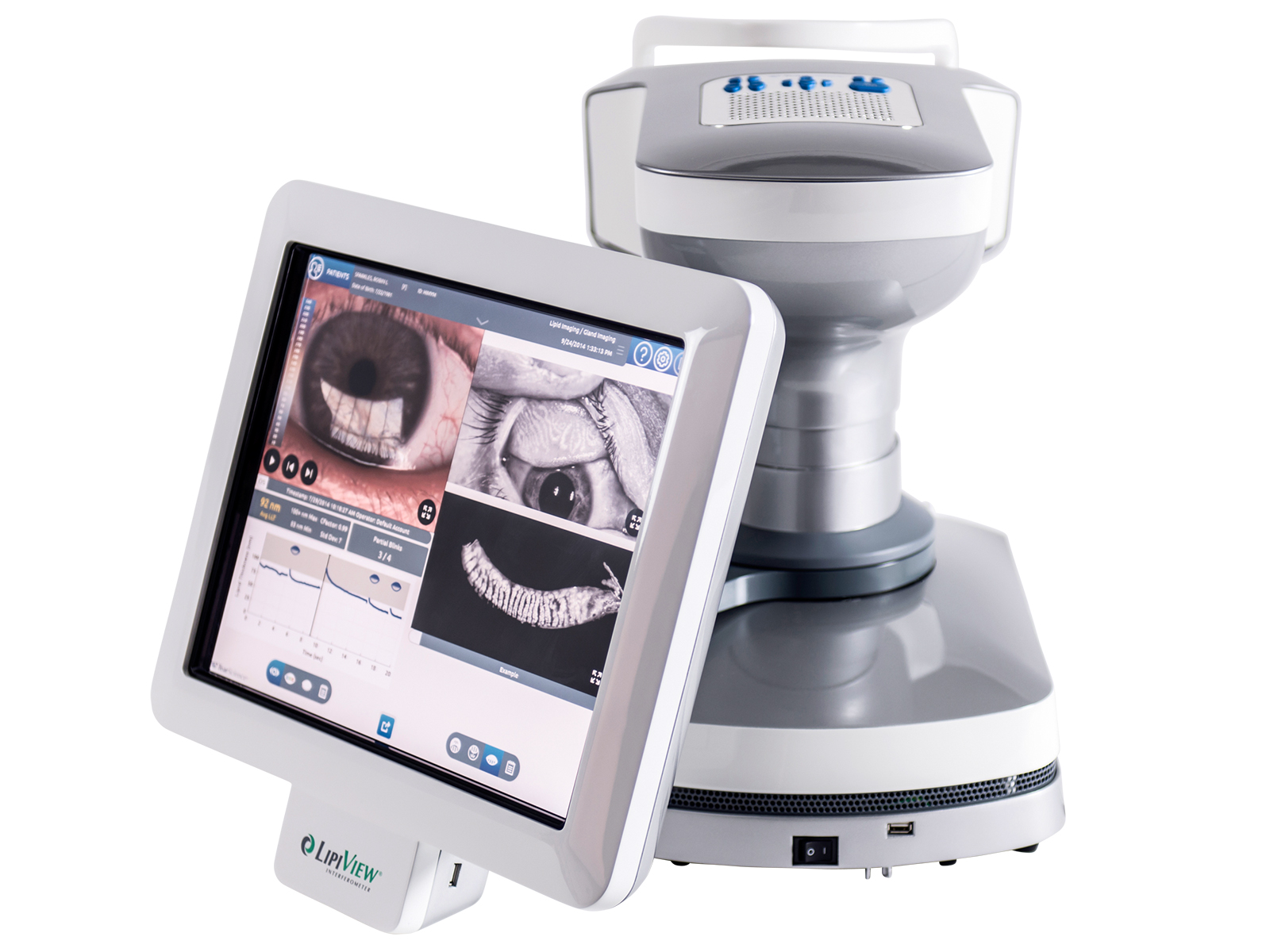
My colleagues and I have been utilizing the LipiView and LipiFlow technology from TearScience for the past several years. The technology is extremely useful, especially the LipiView II with Dynamic Meibomian Imaging which allows us to assess meibomian gland structure and function.
We have all patients fill out a Standard Patient Evaluation of Eye Dryness (SPEED) questionnaire which evaluates the frequency and severity of symptoms. The results range from zero to 28, zero being no symptoms and 28 being intolerable symptoms. We then perform meibography on anyone who scores a 6 or above. The pictures from the LipiView II are obtained quickly and provide a great teaching tool for patients to see and understand their disease process.
We oftentimes have patient who score 6 or above come back to our dry eye clinic where we perform a complete LipiView analysis. We evaluate the oil composition and the patient’s blinking pattern. We then repeat the meibography and do a more thorough analysis of the patients’ overall ocular surface health. That evaluation involves additional testing, meibomian gland evaluation, fluorescein staining, and other, typical dry eye diagnostics.
In my experience, meibography is the most helpful ocular surface evaluation that we perform. The results from some other tests, such as a lipid analysis, can vary if patients applied drops, makeup, or lotion earlier in the day. Meibography, on the other hand, shows gland loss, atrophy, and truncation, and serves to highlight the disease progression that will cause patients to have more symptoms over time.
When incorporating LipiView into your practice, it is important to know that the device needs its own room. Although it takes up some space, it is not more than that of a perimeter or other diagnostic devices that you would have in the office. TearScience was great in training our staff and physicians, and easing us through the learning curve.