- Product Listings
- Compare Products
- Calculators
- Product Tours
- Expert Reviews
- Our Experts
- Company Directory
- About Beye
- Contact Us
Ⓒ 2026 Beye.com. All rights reserved.
This content is intended for health care professionals and providers only. The information contained on Beye.com, including text, graphics, images, and interactive activities, is for informational purposes only, and is not intended to be a substitute for professional medical advice. Beye LLC, via its Editors and Publisher, accepts no responsibility for any injury or damage to persons or property occasioned through the implementation of any ideas or use of any product described herein. Although great care is taken to ensure that all information is accurate, it is recommended that readers seek independent verification of advice on drugs and other product usage, surgical techniques and clinical processes prior to their use. References made in article may indicate usage of medical equipment or drugs at dosages, for periods of time, and in combination not included in the current prescribing information. Inclusion of advertising materials on the website thereof, does not constitute and representation or guarantee by Beye LLC of the quality of such products, or of the claims made.
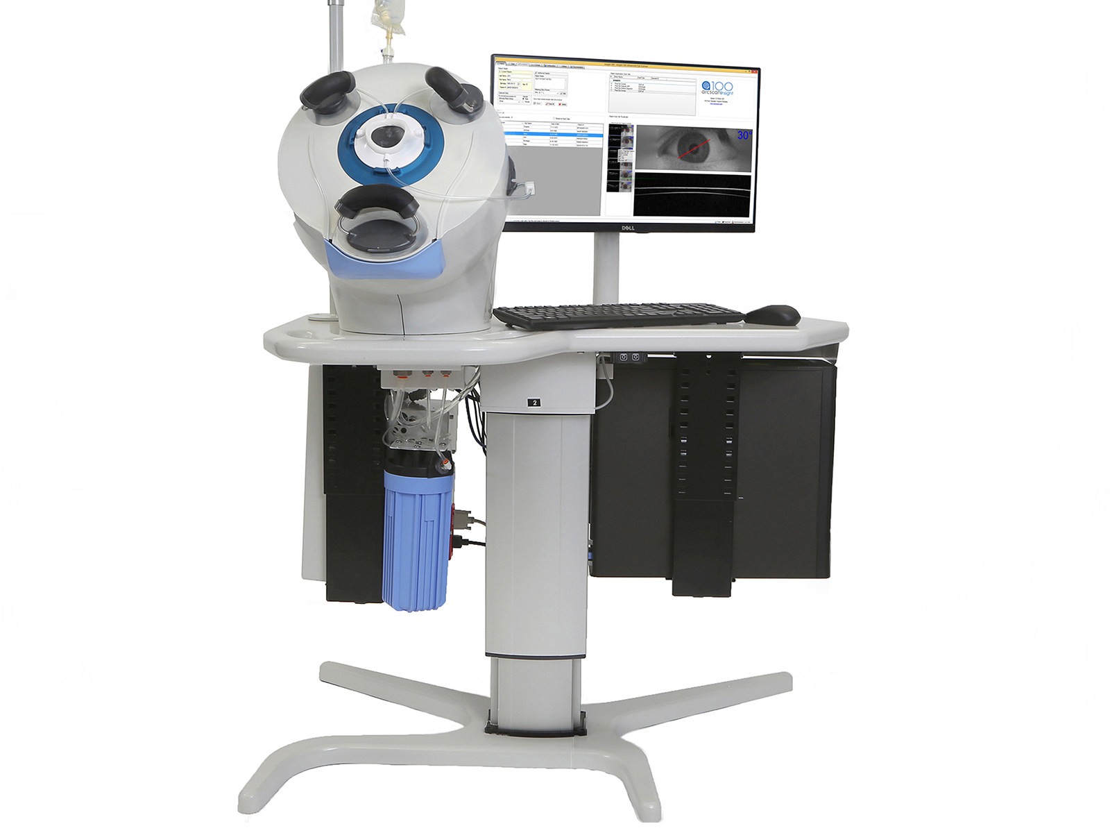
ArcScan Insight 100
ArcScan, Inc.The ArcScan Insight 100 System provides a complete visualization of the true anatomy of the entire anterior segment, including areas behind the iris, and enable precision analysis with repeatable and reproducible data.
At-a-Glance
- Display screen is a monitor
- Software included is ArcScan Insight 100 Software
- Scan Modes: Auto-centering, No centering/manual centering, Continuous scan, Next/repeat scan mode
- Scan Type: Anterior Segment Imaging and Biometry, Capsule Imaging, Capsule Imaging and Biometry
- Measurements: Cornea, Corneal Diameter, Anterior Chamber Depth, Angle-to-Angle width, Sulcus-to-Sulcus width, Pathologic Structures
- Produces images with 1 micron resoultion of the cornea or the anterior segment
Details
FDA
Yes
CE Mark
Yes
Modes
Not specified
System
Standalone Unit
Type
High Frequency
From Your Peers
Show More
User Ratings + Reviews
The Insight 100 is versatile and automated, which simplifies the process of obtaining images. When capturing the images the patient sits up in a chair, then leans forward into a self-contained, sealed water bath for the eye. This process is clean, noninvasive, and easy for physicians and technicians to perform. The high frequency ultrasound allows the Insight 100 to visualize anatomy that OCT can not - this promotes effective lens positioning allowing for more precise power selection. The Insight 100 also provides reliable data for ICL sizing and screening patients with suspect topography for keratoconus using epithelial mapping. And finally using the anterior segment ultrasound we can image behind an inlay for corneal inlay adjustments.
The Insight 100 is a truly innovative technology with wide application. The Insight 100 is unique among ophthalmic imaging devices in the precision and detail of its epithelial mapping and flap thickness measurements for patients with previous LASIK, in its overview of anterior segment shape, measurement of sulcus diameter for use with phakic IOLs, and ability to locate cataracts and zonular attachments for understanding effective lens position after surgery. It offers powerful diagnostic applications, such as screening for keratoconus, understanding epithelial shape before and after corneal cross-linking, determining corneal shape and thickness for patients undergoing INTACS, and measuring inlay depth for patients undergoing KAMRA procedures. Unlike OCT, ArcScan's very high frequency ultrasound technology can see through opacities and image behind the iris.
Show More
Company Information
Contact the company for additional information, availability, or pricing:
ArcScan, Inc.
arcscan.com433 Park Point Drive, Suite 220
Golden, CO 80401

Trending in Diagnostic Instruments
Powered by:
Tracking the Alignment of Toric IOLs
MillennialEye, May/June '18
Pseudophakic Bullous Keratopathy
William B. Trattler, MD
CRSToday, April 2018
Spotting AMD in the OD’s Office
CollaborativeEye, Mar/Apr '18
Spotting AMD in the OD’s Office
Nicole Stout, OD, FAAO, and Nate R. Lighthizer, OD, FAAO
CollaborativeEye, Mar/Apr '18
Talking Torics
CRSToday, March 2018
Cataract Surgery After LASIK Corneal Flap Removal
Karl G. Stonecipher, MD
CRSToday, March 2018
IOP After Short-Term Scleral Lens Wear in Healthy Adults
CollaborativeEye, Debut Issue
Ten Do’s and Don’ts for Cataract Comanagement
CollaborativeEye, Debut Issue
Persistent LASIK Flap Interface Fluid After DSAEK Procedure
CRSToday, February 2018
Persistent LASIK Flap Interface Fluid After DSAEK Procedure
Mark S. Gorovoy, MD
CRSToday, February 2018
Getting Another Shot
William B. Trattler, MD; Alan Berg, MD; and Naja Chisty, DO
CRSToday, January 2018
Dual Benefits of CXL
CRSTodayEurope, Mar 2013
Dual Benefits of CXL
CRSTodayEurope, Mar 2013
My Algorithm for DED
CRSToday, Jan 2016
Show More
