Editorially Independent Content
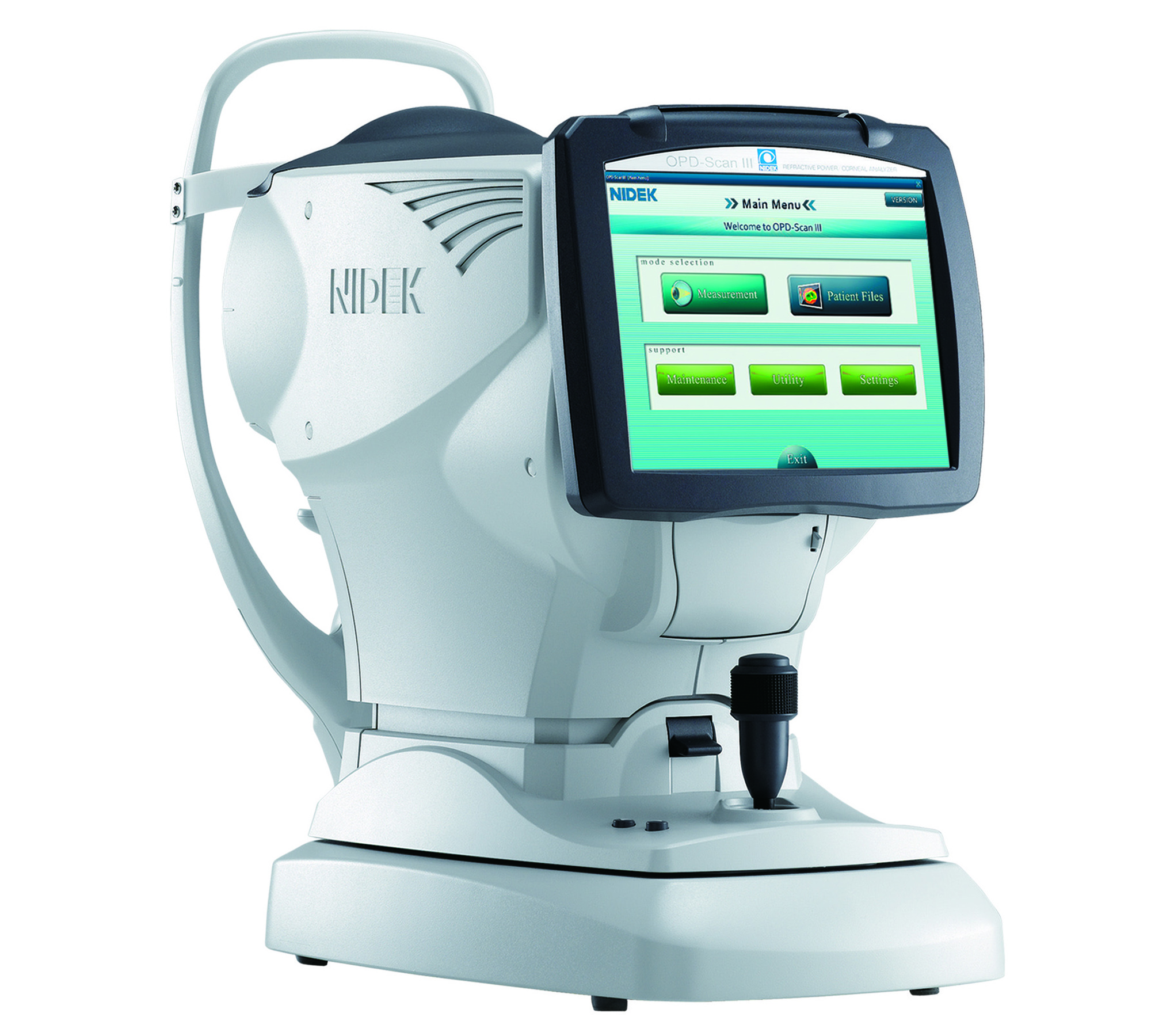
The Nidek OPD III (Nidek, Inc.) is an advanced vision assessment system that captures valuable data about the patient’s eye within 12 seconds. Rather than moving from one instrument to another, the patient can remain seated in one chair for increased office efficiency and patient comfort. The OPD III automatically captures the data from one eye while the patient stares at a light and then, it’s moved over to capture data from the other eye. The system assesses the degree and pattern of astigmatism, identifies corneal surface issues, and gathers data regarding higher order aberrations, mesopic and photopic pupil size and Angle Kappa; information that is vital for determining the best implant option for each patient.
ASSESSING PATIENT NEEDS
My practice has used the Nidek OPD III for quite some time—even dating back to the time when it was known as the 3D-Wave. The OPD III is necessary to calculate and customize the best implant for each eye of every patient. Without it, there is no way to know whether the patient is a candidate for a certain type of implant.
One of the nice features of the OPD III is how easy it is for our technicians to learn to use this device; moreover, the patients are happy because they don’t have to sit for a prolonged period of time with a light shining in their eyes—it captures a very large amount of data extraordinarily quickly.
PREOPERATIVE EVALUATION
The first thing I look at is the root mean square or RMS value. This tells me how clean the image of the light is going in and out of the eye. If the RMS value is high, that means the patient’s postoperative outcome will probably not be 20/20 due to an underlying pathology, such as macular or corneal issues. A high RMS number allows the surgeon to set proper expectations for the patient.
The next thing I look at is the axial map, which tells me the amount, location and pattern of the anterior corneal astigmatism. If it’s an irregular pattern, such as keratoconus or pellucid, then that patient might not be an ideal candidate for a toric implant. However, if it’s a beautiful bowtie pattern, then that patient is a perfect candidate for a toric implant. By showing the bowtie image on a large monitor on the fabulous color maps captured by the OPD III—patients can readily ‘see’ their astigmatism. Therefore, they are more apt to recognize and justify the out-of-pocket cost of a toric implant.
VALUABLE DATA
The other way I use the OPD III is to look at the point spread function. I first show my patients what their total corneal aberration looks like with the cataract in the eye. Then I show them what the image is going to look like after their cataract (the lower aberration) is removed, leaving behind their higher order aberrations. Since some patients have quite a bit of higher order aberration, surgeons can let the patient know ahead of time what to expect after the surgery. For example, a pinpoint of light may look like it has feathered edges or tails. The patients then understand that this is due to their preexisting higher order aberration and not a flaw of the implant or a complication of the surgery.
FOCUSED EXPECTATIONS
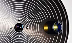
Lastly, for every patient who is undergoing cataract surgery, I use the OPD III to take a Placido disk image of the anterior surface of their cornea and tear film. If the mires are warped, wobbly or irregular in width, rather than concentric and perfectly round, that means the patient has preexisting ocular surface disease. Depending on its severity, I may choose to postpone the cataract surgery in order to rehabilitate the ocular surface to obtain more accurate pre-op measurements.
Also, by showing patients the irregular mires on the Placido disk image, they realize that they have preexisting dry eye syndrome instead of blaming their surgeon for causing their post operative Dye Eye condition.
There is a bit of a challenge for the surgeon to learn how to decipher all of the maps. Marco Ophthalmic, Inc. offers information on its website to help surgeons learn how to interpret the maps and the Marco Reps are more than happy to help surgeons understand how to best utilize the information generated from the various map.