- Product Listings
- Compare Products
- Calculators
- Product Tours
- Expert Reviews
- Our Experts
- Company Directory
- About Beye
- Contact Us
Ⓒ 2026 Beye.com. All rights reserved.
This content is intended for health care professionals and providers only. The information contained on Beye.com, including text, graphics, images, and interactive activities, is for informational purposes only, and is not intended to be a substitute for professional medical advice. Beye LLC, via its Editors and Publisher, accepts no responsibility for any injury or damage to persons or property occasioned through the implementation of any ideas or use of any product described herein. Although great care is taken to ensure that all information is accurate, it is recommended that readers seek independent verification of advice on drugs and other product usage, surgical techniques and clinical processes prior to their use. References made in article may indicate usage of medical equipment or drugs at dosages, for periods of time, and in combination not included in the current prescribing information. Inclusion of advertising materials on the website thereof, does not constitute and representation or guarantee by Beye LLC of the quality of such products, or of the claims made.
Compare UBM
9 Products
reset all
At-a-Glance
Description
FDA
CE Mark
Modes
System
Type
- Display screen is 10.1 in. high-resolution multi-touch monitor (1280 x 800 pixel)
- System Network: Data Archive/Export Capability (one touch export images (.jpg), video clips (.AVI), and exam reports (.PDF) for referral, presentation, or EMR), EHR, Ethernet Printer (any windows printer), Bluetooth 4.0, 802.11n dual-band Wi-Fi and USB 3.0 Ports, Operating system is Windows
- Adjustable angle kickstand and VESA bracket for articulating arm or wall mounting
The VuPad is a portable device that delivers exceptional image quality in a wide range of applications. It is easy to use, and allows the user to capture still images and video.
Yes
Not specified
Not specified
Standalone Unit
A/B-Scan/UBM

- Display screen is a large 24 in. High-Resolution Monitor (1920 x 1080 pixel)
- Size is Large 24 in. High-Resolution Monitor (1920 x 1080 pixel) and weight is 13 lbs. (console only)
- System Network: Data Archive/Export Capability, VGA, HDMI, 5 USB Ports, Bluetooth (for foot pedal, keyboard, and mouse), Ethernet, Operating System is Windows 8, Printer (any Windows-compatible printer)
- Easy interface with image management and EMR systems
- Capture 50-frame video clips at up to 20 frames per second
The VuMax HD offers the ability to be configured with probe B, UBM or an optional combination of probe A. The VuMAX HD also features a database to search, and enhanced focus rendering.
Yes
Not specified
Not specified
Standalone Unit
UBM/B-Scan
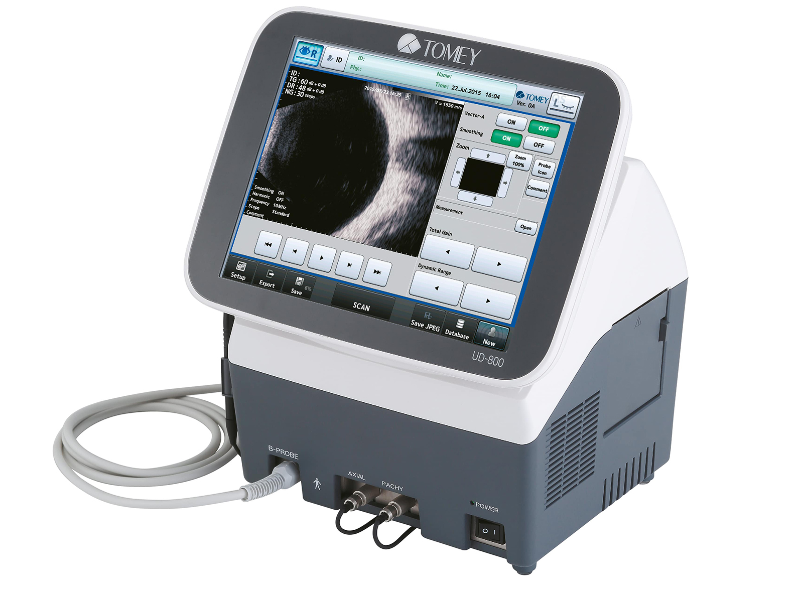
- Size is 310 (W) x 214 (D) x 326 (H) mm and weight is 6.0 kg
- Display screen is a TFT color LCD touchscreen with a 10.4" screen
- Measurements: Anterior Chamber Depth, Lens Thickness, Pachymetry
- IOL Calculation: HAIGIS, HOFFER-Q, HOLLADAY I, SRK-T, SRK II
- System Network: Data Archive/Export Capability, USB, Printer (networked or local)
The Ultrasonic A/B Scanner and Pachymeter UD-800 is compact all-in-one device with high resolution. Main mode is B-Scan, and you can add UBM, Biometry, Pachymetry and A-Scan Diagnosis as options. The calculation system is same as OA-2000 and EM-4000, which makes it easy for users to operate. The UD-800 can save data up to 52,000 eyes (B-Scan) and print out the data with the built-in printer. Also Harmonic function is available with B-Scan.
Not specified
Yes
Not specified
Standalone Unit
A/B-Scan/UBM
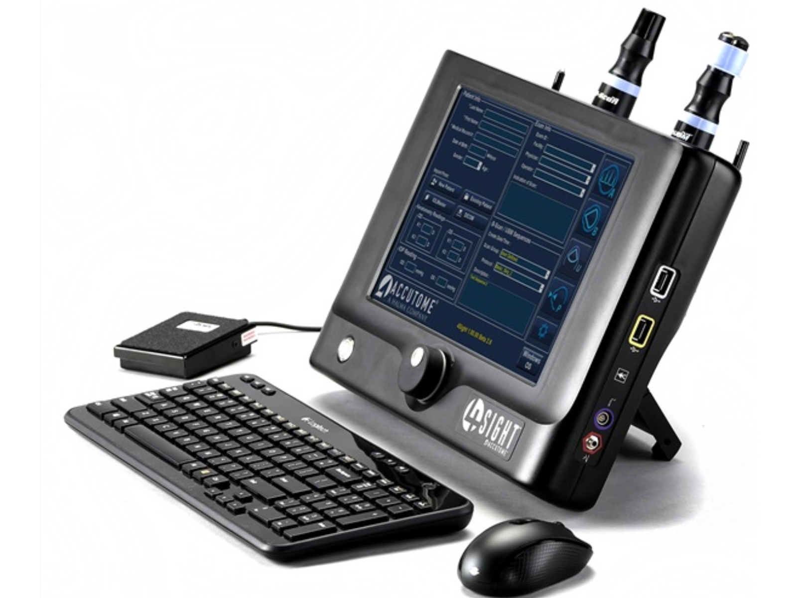
- Clinical accuracy is 0.1 mm and electronic accuracy is 0.016 mm
- Size is 15”W x 2”D x 11”H and weight is 5 lbs
- Display screen is a touchscreen
- Measurements: Anterior Chamber Depth, Lens Thickness, Pachymetry
- IOL Calculation: BINKHORST-II, HAIGIS, HOFFER-Q, HOLLADAY I, SRK-T, SRK II
- System Network: Data Archive/Export Capability, USB, DICOM
- Proprietary signal processing
Designed with eye care professionals in mind, the 4Sight provides a single solution for ophthalmic diagnostics by combining an A-Scan, B-Scan, UBM and Pachymeter in one, easy to use platform.
Yes
Yes
Cataract, Dense Cataract, Aphakic, Pseudophakic, Custom eye types possible
Standalone Unit
A/B-Scan/UBM
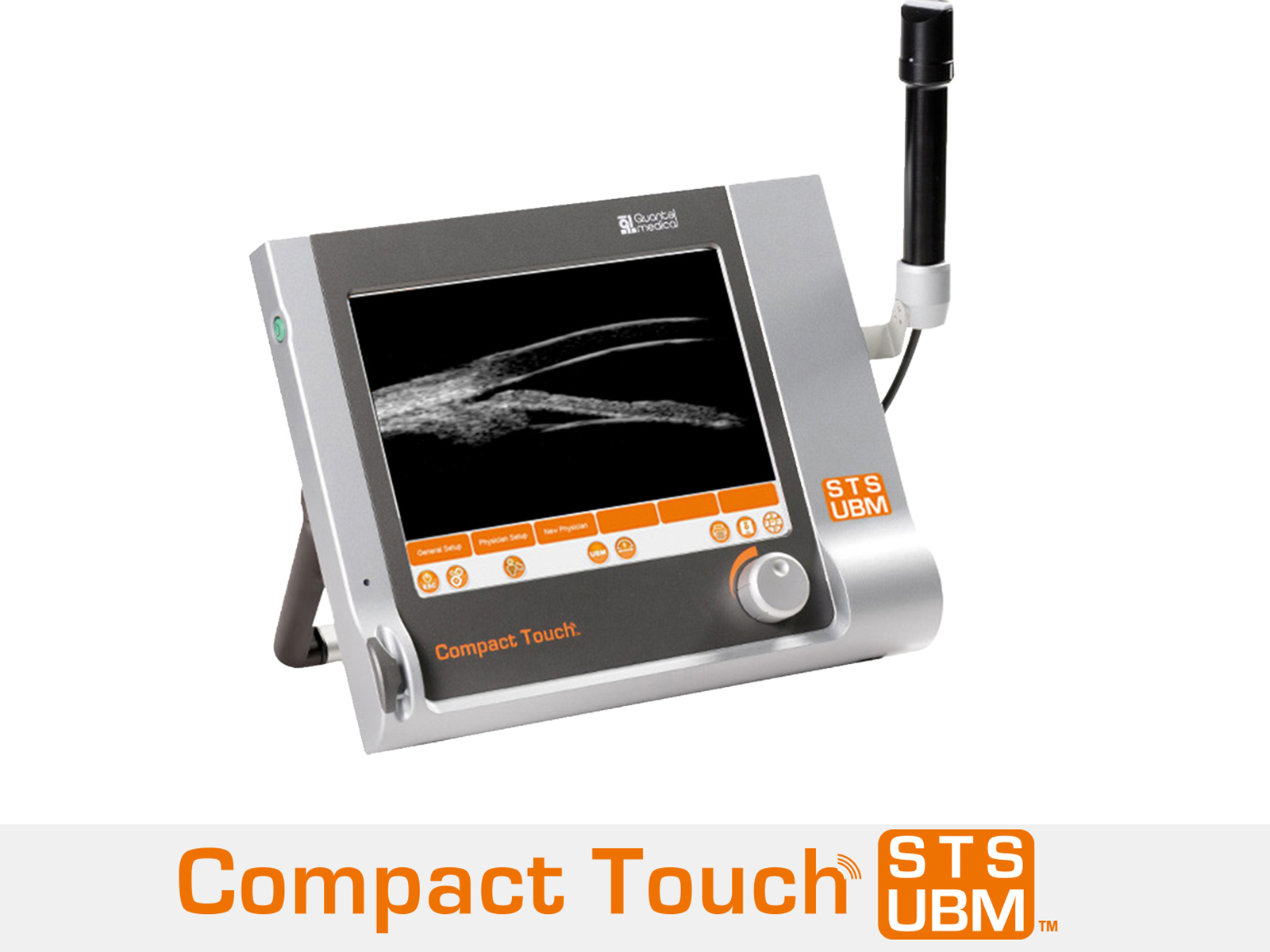
- Adjustable Gain is 20 to 110 dB and Time Gain Control (TGC) is 0 to 30 dB
- Size is 37.5 cm (w) x 10 cm d) x 27 cm (h) - 14.76 in. (w) x 3.94in. (d) x 10.63 in. (h) and weight is 4.28 Kg - 9.26 lb.
- Display Screen is 10.4 in. - 21 cm (w) x 16 cm (h) - 8.3 in. (w) x 6.3 in. (h)
- System Network: Data Archive/Export Capability, USB, Ethernet, EHR, Operating System is Windows 8-Windows 7, Printer (external PC printer Windows 7 (32 bits) compatible)
- Optional STS module for sulcus-to-sulcus measurement and visualization of phakic IOL implantation
- Large range of applications: glaucoma, cataract and refractive surgery
Compact Touch STS UBM is a portable ultrasonic biomicroscope (UBM) for imaging the eye from the cornea to the lens. In comparison to optical technologies, ultrasound can travel through opaque media & pigmented tissues to produce high quality images of ocular structures, especially behind the iris.
Yes
Yes
Not specified
Standalone Unit
UBM
- Electronic accuracy is 0.04 mm - 0.002 in.
- Size is 9 cm (l) x 17 cm (w) x 19 cm (h) - 7.5 in. (l) x 6.7 in. (w) x 7.5 in. (h) and weight is 1.5 kg - 3.3 lbs.
- DIsplay screen is a touchscreen that is 8.6 cm (w) x 11.5 cm (h) - 3.4 in. (w) x 4.5 in. (h)
- Measurements: Anterior Chamber Depth, Lens Thickness
- IOL Calculation: BINKHORST-II, HAIGIS, HOFFER-Q, HOLLADAY I, SRK-T, SRK II. Post Refractive Formula Integration (Pre-op & Post-op Keratometry 6 different methods for keratometric correction and implant calculation: History Derived Refraction Derived Contact Lens Method ROSA Regression SHAMMAS Regression Double K/SRK-T (Dr. ARAMBERRI's formula))
- System Network: Data Archive/Export Capability, USB, DICOM, EHR, Printer (networked or local), Operating System is Windows
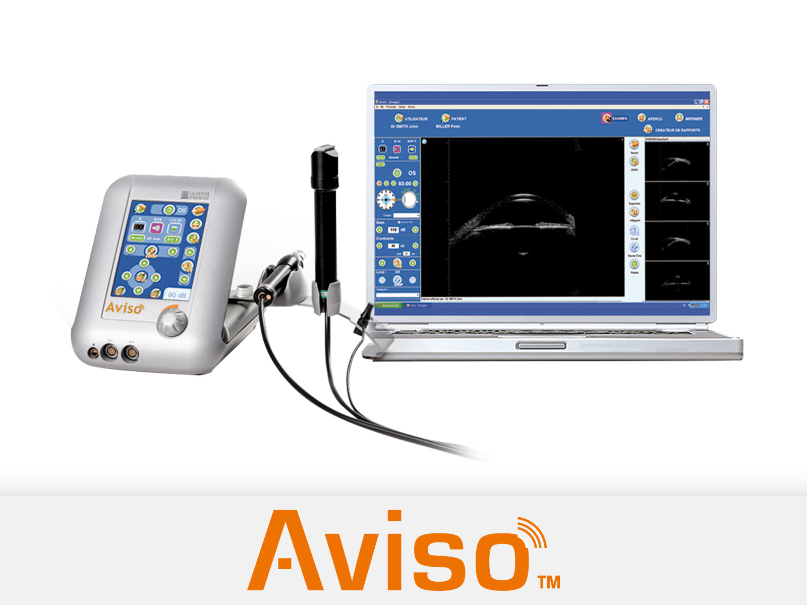
Flagship ultrasound system from Quantel Medical, Aviso offers the largest choice of probe frequencies in the market: 10 MHz, 20 MHz high frequency for posterior segment, UBM (25 MHz and 50 MHz) for the anterior segment, with also biometry and biometry in B mode. Also, the powerful and user friendly interface has a Glaucoma module and Varigain and Cineloop modes.
Yes
Yes
Aphakic, Pseudophakic, Manual, Built-in pattern recognition: PMMA, Acrylic & Silicone Material for Pseudo-phakic Eye types, Selectable Tissue Velocity
Laptop Based
A/B-Scan/UBM
- Electronic accuracy is 0.04 mm - 0.002 in.
- Size is 19 cm (l) x 17 cm (w) x 19 cm (h) - 7.5 in. (l) x 6.7 in. (w) x 7.5 in. (h) and weight is 1.5 kg - 3.3 lbs.
- Display screen is 8.6 cm (w) x 11.5 cm (h) - 3.4 in. (w) x 4.5 in. (h) touchscreen
- Measurements: Anterior Chamber Depth, Lens Thickness
- IOL Calculation: HOFFER-Q, HOLLADAY I, SRK-T, SRK II, Post-Refractive Formulas (Pre-op & Post-op Keratometry 6 different methods for keratometric correction and implant calculation: History Derived Refraction Derived Contact Lens Method ROSA Regression SHAMMAS Regression Double K/SRK-T (Dr. ARAMBERRI's formula))
- System Network: Data Archive/Export Capability, Ethernet, USB, DICOM, EHR, Printer (networked or local), Operating System is Windows
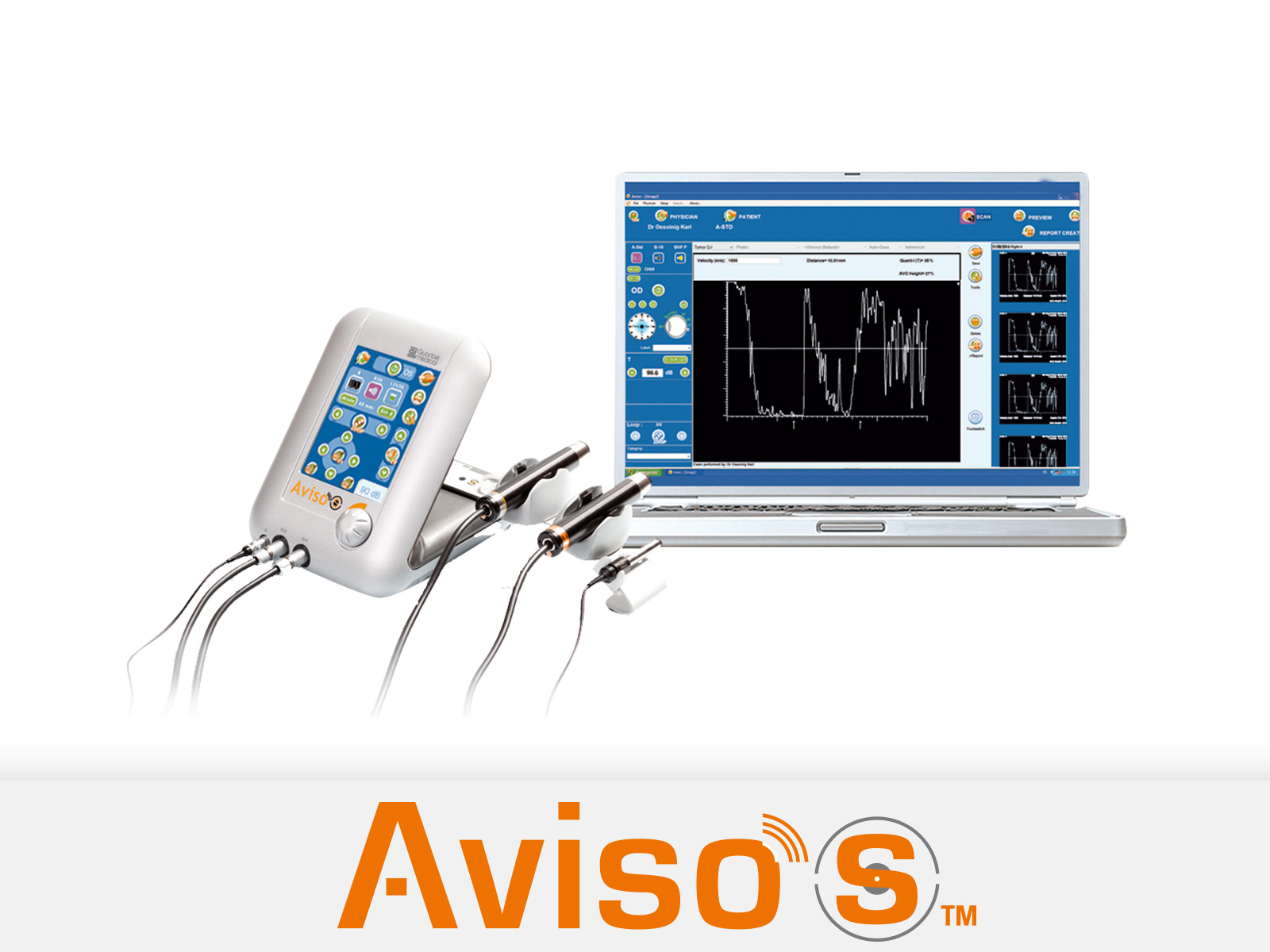
Aviso S is the only platform available in the market to fully comply with the requirements set by Prof. K. Ossoinig to conduct standardized echography. Aviso S offers 10 MHz, 20 MHz high frequency , UBM (25 MHz - 50 MHz), biometry, biometry in B mode and A Standardized probe.
Yes
Yes
Aphakic, Pseudophakic, Manual, Built-in pattern recognition: PMMA, Acrylic & Silicone Material for Pseudo-phakic Eye types, Selectable Tissue Velocity
Laptop Based
A/B-Scan/UBM
- Adjustable Gain is 0 - 112 dB and Time Gain Control is near, middle, far
- Display Screen is 15.4 inch WSXGA+ LCD or larger
- System Network: Data Archive/Export Capability, EHR, DICOM, Operating System is Windows XP, Windows Vista, WIndows 7, WIndows 8, Printer (networked or local)
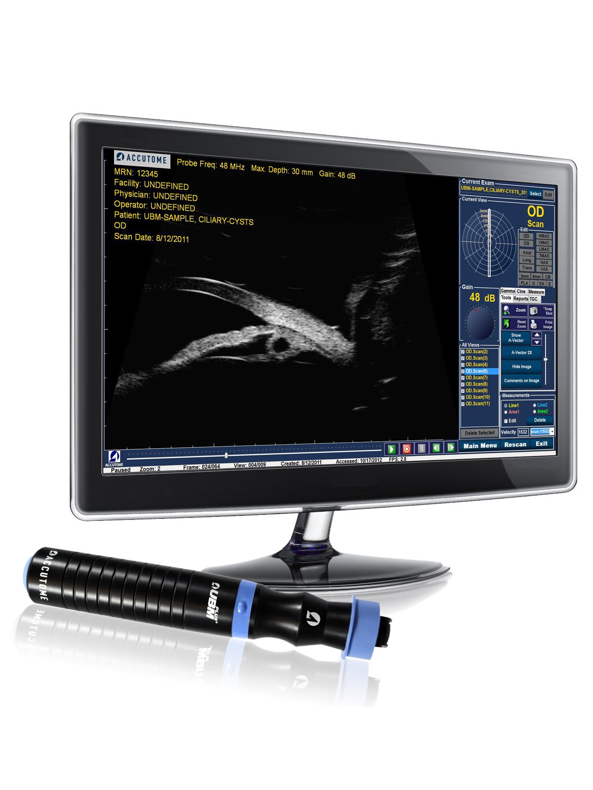
- Contains measuring tools along with corresponding A-Vector. Useful for measuring sulcus-to-sulcus, anterior chamber depth, positioning of intraocular lenses and filtration angle of the eye
- Detailed structure definition of the anterior segment of the eye
Accutome's UBM Plus is used for anterior segment imaging. The UBM Plus is an incredibly portable device because the 48 MHz probe plugs directly into a laptop or desktop computer. This unit features an all-in-one probe design to eliminate signal loss and provide sharpest images possible.
Yes
Yes
Not specified
Laptop Based
UBM
- Size with stand is: 15 inches (390 mm) W x 15.5 - 20 inches (400 - 520 mm) H x 10 inches (260 mm) D size without stand: 17.5 in. (h) x 17 in. (w) x 10.5 in. (d) and weight is 23.5 lbs. (10.75 kg)
- Unique touch screen display allows for input without the need for a keyboard (keyboard/mouse are included)
- System Network: Data Archive/Export Capability, USB, DICOM
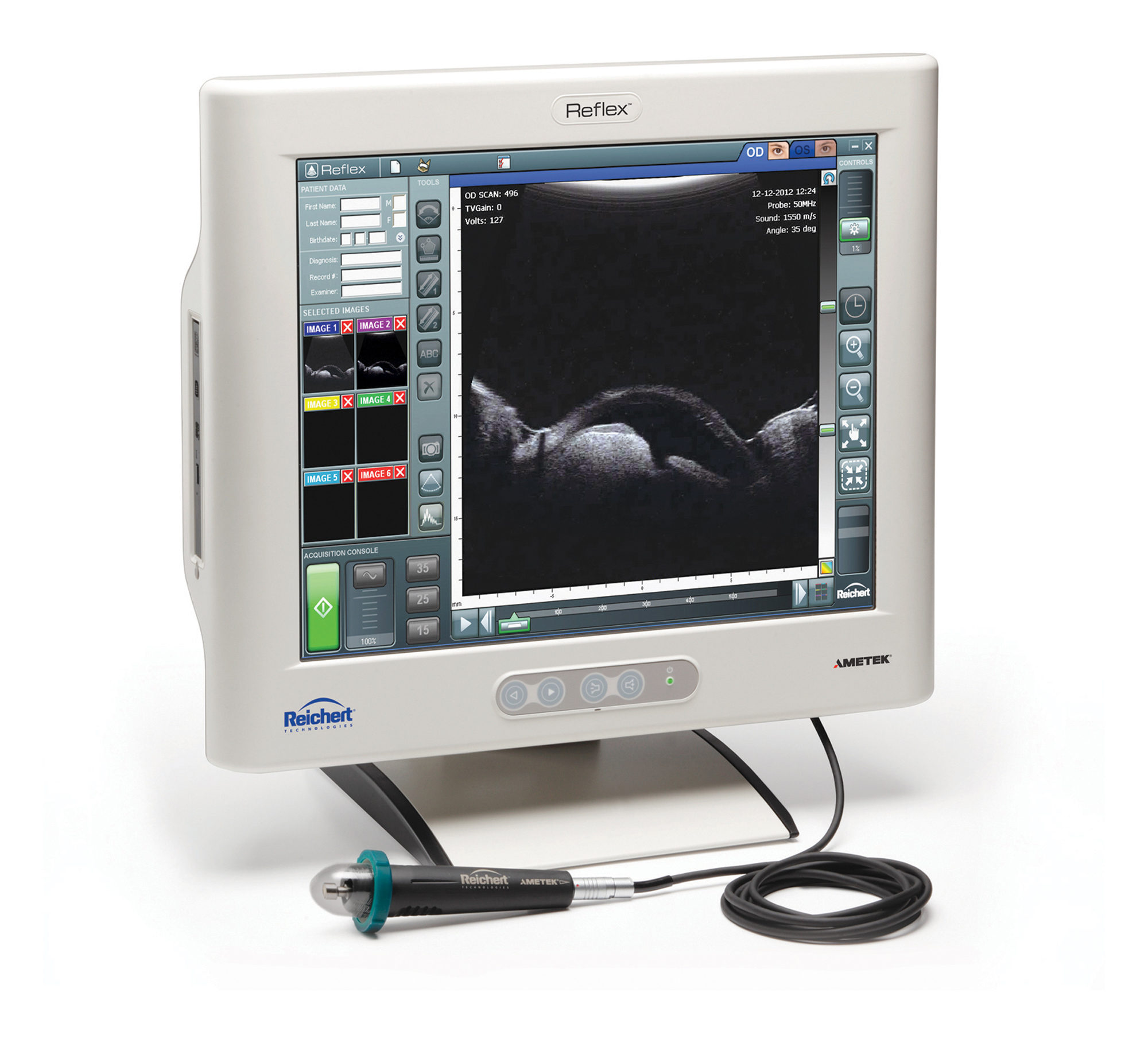
- Specific features in an image may be evaluated using angle, area, and ruler analysis tools
- SThe Reichert Reflex Ultrasound Biomicroscope is a compact, all-in-one, ultrasound instrument for imaging the anterior segment of the eye. Users can now image pathologies, which may be obstructed by opacities or ocular structures, that OCT technology cannot.
Yes
Yes
Not specified
Standalone Unit
UBM
Privacy | Terms & Conditions Ⓒ 2026 Beye, LLC. All rights reserved.
